


If you have ever received an ultrasound report and wondered what words such as echogenicity, hypoechoic, calculus, cyst, normal size or no focal lesion actually mean, you are not alone. An ultrasound report can look complicated, even when the findings are completely normal.
USG, or ultrasonography, is one of the most commonly used medical imaging tests. It helps doctors look at organs, tissues, blood vessels and, during pregnancy, the developing baby. Unlike X-rays and CT scans, ultrasound uses sound waves rather than ionising radiation.
The important thing to remember is that an ultrasound report is not designed to be read like a simple pass-or-fail test. It contains measurements, observations and medical terminology that need to be understood in the context of your symptoms, examination and other investigations.
This guide explains what USG means, what an ultrasound can show, the different types of USG scans and, most importantly, how to approach your USG report without unnecessarily worrying about every unfamiliar word.
What does USG mean?
USG stands for ultrasonography. It is a medical imaging technique that uses high-frequency sound waves to create images of structures inside the body.
During a USG scan, a device called a transducer or probe sends sound waves into the body. These waves bounce back differently depending on the tissues and organs they encounter. The ultrasound machine processes these returning echoes and produces images that can be viewed in real time.
The terms USG, ultrasound, ultrasonography and sonography are commonly used to describe the same basic imaging technology.
A USG report, however, is different from the scan itself. The scan produces images, while the USG report is the written interpretation of those images, usually prepared by a radiologist or other appropriately trained medical professional.
What is a USG test?
A USG test is a non-invasive imaging examination used to examine organs, tissues and blood flow inside the body.
Depending on the type of scan, ultrasound may be used to examine the:
- Liver
- Gallbladder
- Kidneys
- Urinary bladder
- Pancreas
- Spleen
- Thyroid gland
- Uterus and ovaries
- Prostate
- Breasts
- Blood vessels
- Muscles, tendons and soft tissues
- Developing fetus during pregnancy
Doctors may recommend a USG when someone has abdominal pain, pelvic pain, swelling, unexplained bleeding or other symptoms. It can also be used to monitor known conditions, assess pregnancy and guide certain procedures such as needle biopsies.
What does an USG scan show?
An ultrasound scan can provide information about the size, shape, position and appearance of organs and tissues.
For example, an abdominal USG may help identify gallstones, kidney stones, cysts, fluid collections, enlargement of certain organs or other structural abnormalities. A thyroid ultrasound can examine nodules and changes in the thyroid gland. A pelvic ultrasound can assess the uterus and ovaries.
Ultrasound can also distinguish certain types of structures. For example, it may help doctors differentiate a fluid-filled cyst from a solid mass. However, ultrasound cannot always determine exactly what a mass is, and further testing may sometimes be required.
Doppler ultrasound adds another important capability. It can assess the movement of blood through blood vessels, including the direction and speed of blood flow.
This is why an ultrasound report should not be interpreted based on one word or measurement alone. The complete picture matters.
Is ultrasound and USG the same?
Yes. Ultrasound and USG generally refer to the same imaging technology.
USG is short for ultrasonography, while ultrasound is the more commonly used term. You may also see terms such as sonography or sonogram.
There can be differences in the specific examination performed—for example, an abdominal ultrasound, pelvic USG or Doppler USG—but they all use ultrasound technology.
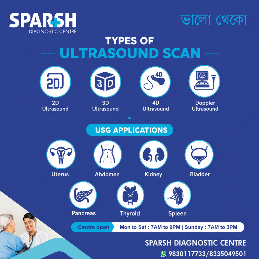
Types of USG scans
There is no single type of ultrasound scan. The examination is selected according to the body part being assessed and the medical question the doctor wants answered.
2D ultrasound
2D ultrasound is the standard and most commonly used form of ultrasound imaging. It produces two-dimensional images of organs and tissues and is widely used for routine diagnostic examinations and pregnancy scans.
3D ultrasound
3D ultrasound creates three-dimensional images by collecting information from different angles. It can provide additional visual detail in certain examinations.
4D ultrasound
4D ultrasound is essentially 3D ultrasound displayed in motion. It can produce moving images in real time and is particularly familiar in pregnancy imaging. However, 3D and 4D imaging do not replace medically necessary diagnostic assessment.
Doppler ultrasound
Doppler ultrasound evaluates blood flow. It can be used to examine arteries and veins and may help identify problems such as narrowing, blockage or abnormal blood flow. In pregnancy, Doppler may be used when assessment of fetal or placental blood flow is clinically indicated.
Abdominal ultrasound
An abdominal USG examines organs such as the liver, gallbladder, pancreas, kidneys, spleen and urinary bladder.
It may be recommended for abdominal pain, suspected gallstones, kidney stones, abnormal liver findings and several other conditions.
Pelvic ultrasound
Pelvic USG evaluates structures within the pelvis. Depending on the patient, it may examine the uterus, ovaries, urinary bladder and other pelvic structures.
Obstetric ultrasound
Obstetric ultrasound is performed during pregnancy to monitor fetal growth and development, assess fetal anatomy, examine the placenta and amniotic fluid, and evaluate other aspects of pregnancy when clinically indicated.
Transvaginal ultrasound
A transvaginal ultrasound uses a specially designed probe inserted into the vagina to obtain clearer images of the uterus and ovaries. It is commonly used in early pregnancy, infertility evaluation and the assessment of pelvic symptoms.
Thyroid ultrasound
A thyroid USG examines the thyroid gland and can help identify nodules, cysts, enlargement and other structural changes.
Breast ultrasound
Breast ultrasound can be used to evaluate a breast lump or other breast symptoms. It can help determine whether a finding appears fluid-filled or solid, although additional tests may be needed depending on the finding.
Musculoskeletal ultrasound
This type of ultrasound examines muscles, tendons, ligaments, joints and other soft tissues. It can be useful for evaluating certain sports injuries, tendon problems and soft-tissue abnormalities.
How to read your USG report
Reading an ultrasound report does not mean trying to diagnose yourself. Instead, think of it as learning what each section means before discussing the findings with your doctor.
Most reports contain several basic components.
1. Check the type of examination
Start at the top of the report and identify exactly what scan was performed.
For example, it may say:
- USG whole abdomen
- USG abdomen and pelvis
- Pelvic USG
- Obstetric USG
- Thyroid ultrasound
- Breast ultrasound
- Doppler study
This matters because the expected findings and measurements vary according to the type of examination.
2. Look at the organs examined
The report will usually discuss each organ separately.
For example, an abdominal ultrasound may contain sections for the liver, gallbladder, pancreas, spleen, kidneys and urinary bladder.
The radiologist may describe the size, shape, position, texture and appearance of each structure.
3. Understand measurements
Ultrasound reports often contain measurements in centimetres or millimetres.
For example, you may see the length of a kidney, the size of the liver, the thickness of the endometrium or the dimensions of a cyst.
Do not assume that a number outside your expectations is automatically abnormal. Normal measurements vary depending on the organ, age, sex, pregnancy status and clinical situation.
The reference values used by the radiologist and the overall appearance of the structure are more important than a number viewed in isolation.
4. Understand common words
Some ultrasound terminology appears frequently.
Normal size: The organ’s size is within the expected range.
No focal lesion: No specific localised abnormality was identified in the examined area.
Cyst: A fluid-filled structure. Many cysts are benign, but their significance depends on their location, appearance and size.
Calculus: A stone. For example, a renal calculus means a kidney stone.
Echogenicity: This describes how tissues reflect ultrasound waves and how they appear on the scan.
Hypoechoic: A structure reflects fewer ultrasound waves than surrounding tissue and therefore appears relatively darker.
Hyperechoic: A structure reflects more ultrasound waves and appears relatively brighter.
No free fluid: No abnormal collection of free fluid was seen in the area examined.
Bilateral: Refers to both sides, such as both kidneys or both ovaries.
Unremarkable: This usually means no significant abnormality was identified in the structure being described.
These words can sound alarming when read without context. In many cases, they are simply standard descriptive terms used by the radiologist.
5. Pay attention to the impression
The Impression or Conclusion is often one of the most useful sections of the report.
It summarises the important findings from the examination.
For example, a report may describe several organs as normal and then provide a short impression mentioning one clinically relevant finding.
This section can help your treating doctor quickly understand the main points of the examination. However, it still needs to be interpreted alongside your symptoms and medical history.
6. Do not panic over one abnormal word
An abnormal ultrasound finding does not automatically mean cancer or another serious disease.
For instance, an ultrasound may identify a simple cyst, mild fatty changes in the liver, a small kidney stone or another relatively common finding.
The significance depends on the complete report, your symptoms, your age, previous investigations and what your doctor is looking for.
Likewise, a completely normal ultrasound does not guarantee that there is no medical problem. Some conditions cannot be seen clearly on ultrasound, and certain symptoms may require blood tests, CT, MRI, endoscopy or other investigations.
Can USG detect gender?
Ultrasound may sometimes show fetal anatomy that can indicate sex during pregnancy, depending on factors such as gestational age, fetal position and image quality.
However, in India, disclosure of the sex of the fetus during prenatal diagnostic procedures is prohibited by law. The Pre-Conception and Pre-Natal Diagnostic Techniques (Prohibition of Sex Selection) Act, 1994 prohibits determination and communication of fetal sex for the purposes addressed by the Act.
The purpose of pregnancy ultrasound is to assess the health and development of the mother and fetus—not to determine fetal sex.
Is ultrasound 100% safe?
No medical procedure should casually be described as “100% safe.” However, diagnostic ultrasound has a strong safety record and is generally considered safe when appropriately performed by trained healthcare professionals.
One major advantage is that ultrasound does not use ionising radiation, unlike X-rays and CT scans. This is one reason it is widely used during pregnancy.
At the same time, ultrasound energy can produce biological effects under certain conditions. Regulatory and professional organisations therefore recommend that ultrasound be used prudently and for appropriate medical reasons rather than for unnecessary exposure.
In simple terms: when your doctor recommends a medically indicated ultrasound and it is performed appropriately, it is generally considered a safe imaging test.
What can affect the accuracy of a USG scan?
Ultrasound is extremely useful, but it has limitations.
Image quality can be affected by:
- Excess intestinal gas
- Body habitus
- The location and depth of an organ
- The patient’s ability to maintain the required position
- Fetal position during pregnancy
- The skill and experience of the operator
- The type of ultrasound equipment used
Some organs and abnormalities are simply easier to see with ultrasound than others.
This is why your doctor may recommend another imaging test if the ultrasound does not provide enough information.
When should you discuss your USG report with a doctor?
You should discuss your report with the doctor who ordered the scan, particularly if the report mentions:
- A mass or suspicious lesion
- A large cyst
- Gallstones or kidney stones
- Enlarged organs
- Abnormal blood flow
- Fluid collections
- Significant changes from a previous scan
- Findings described as requiring correlation or follow-up
Even when the report appears normal, speak with your doctor if your symptoms continue. A normal scan does not necessarily explain every symptom or exclude every possible condition.
Final thoughts
Understanding your USG report can make the experience much less confusing. The key is to look at the report as a whole rather than focusing on one unfamiliar word or measurement.
USG, or ultrasonography, is a versatile imaging test used to examine many organs and tissues, assess blood flow and monitor pregnancy. It is non-invasive, does not use ionising radiation and has an established role in modern diagnosis.
When reading your report, first identify the type of scan, then review the organs examined, measurements and descriptions, and finally look at the impression. Most importantly, remember that an ultrasound report is only one part of a medical evaluation.
If something in your report concerns you, don’t rely on internet searches to diagnose yourself. Your doctor can interpret the findings in the context of your symptoms, examination and other test results and tell you whether you need treatment, monitoring or further investigation.
Frequently asked questions about USG
What is a USG test?
A USG test is an ultrasound imaging examination that uses high-frequency sound waves to create pictures of organs, tissues and other structures inside the body. It is non-invasive and does not use ionising radiation.
What does USG mean?
USG stands for ultrasonography. Ultrasound, ultrasonography and sonography are commonly used terms for this type of medical imaging.
What does an USG scan show?
An USG scan can show the size, shape, position and appearance of many organs and soft tissues. Depending on the type of scan, it can help identify stones, cysts, fluid collections, structural abnormalities and other findings. Doppler ultrasound can also evaluate blood flow.
Is ultrasound and USG the same?
Yes. USG is short for ultrasonography, while ultrasound is the commonly used name for the same basic imaging technology.
Can USG detect gender?
Ultrasound may sometimes show fetal anatomy that can indicate sex, but disclosure of fetal sex during prenatal diagnostic procedures is prohibited in India under the PCPNDT Act. Pregnancy ultrasound is performed for medical assessment of the mother and fetus.
Is ultrasound 100% safe?
Ultrasound is generally considered safe when medically indicated and performed appropriately by trained professionals. It does not use ionising radiation, although ultrasound energy can have biological effects, which is why prudent medical use is recommended.
What are the main types of USG scans?
Common types include 2D, 3D, 4D and Doppler ultrasound, as well as abdominal, pelvic, obstetric, transvaginal, thyroid, breast and musculoskeletal ultrasound. The appropriate examination depends on the patient’s symptoms and the clinical question.
How do I read my USG report?
Start by checking the type of scan and the organs examined. Then look at the measurements and descriptions before reading the Impression or Conclusion section. Avoid interpreting individual terms in isolation. Your doctor should explain whether a finding is significant based on your symptoms and medical history.
Does an abnormal USG report always mean something serious?
No. Ultrasound may identify many benign or insignificant findings. The importance of an abnormality depends on its appearance, size, location, symptoms and other clinical information.
Can a normal USG report rule out every disease?
No. Ultrasound has limitations, and some diseases or abnormalities may not be visible. If symptoms persist despite a normal scan, your doctor may recommend additional evaluation or another type of investigation.
How long does a USG scan take?
The duration depends on the type of examination. Many routine ultrasound examinations can be completed within around 30 minutes to an hour, although the exact time varies.
Do I need to prepare before a USG scan?
Preparation depends on the examination. Some scans require no preparation, while others may require fasting or a full bladder. Your diagnostic centre should tell you exactly how to prepare before the appointment.
Medical disclaimer: This article is intended for general health education and should not be used to diagnose or treat a medical condition. Always discuss your ultrasound findings with a qualified healthcare professional who can interpret the report in the context of your individual health.
#BhaloTheko
Disclaimer:
No content on this site, regardless of date, should ever be used as a substitute for direct medical advice from your doctor or other qualified clinician.




[…] Studies: Includes X-rays, ultrasound, CT scans, or MRI to visualize internal organs and detect issues such as tumors, stones, or […]
[…] Ultrasound […]
[…] Ultrasound – Initial, cost-effective screening tool […]
[…] cases of injury or suspected internal bleeding, imaging such as ultrasound, X-ray, or MRI may be […]
[…] Ultrasound or MRI to assess liver, spleen, and brain involvement […]
[…] CT Scan or MRI: Helpful in assessing spleen size and identifying associated pathologies. […]
[…] Ultrasound abdomen […]
[…] Ultrasound (FAST scan for internal bleeding) […]
[…] Ultrasound – useful for detecting cysts […]
[…] Ultrasound […]
[…] Ultrasound of the abdomen – first-line test […]
[…] scans, MRI, or ultrasound may be used if ischemic colitis, tumors, or structural abnormalities are […]
[…] Ultrasound/CT scan – For suspected amoebic liver abscess […]
[…] Ultrasound to check kidney structure. […]
[…] Ultrasound […]
[…] Contrast-enhanced CT scan: The most common and effective imaging tool for identifying SVT. […]
[…] CT Scan or MRI: Detects tumor mass or liver metastasis […]
[…] Ultrasound: Identifies fluid accumulation or abscesses. […]
[…] Ultrasound Abdomen […]
[…] anomaly scan is a detailed ultrasound examination done in the second trimester of pregnancy, typically between 18 to 22 weeks. Its primary purpose is […]
[…] Ultrasound can visualize nerve compression or structural abnormalities. […]
[…] Tests: Ultrasound, CT scan, or MRI to visualize deep lymph […]
[…] Ultrasound: Often used initially to detect gallstones or ductal obstruction. […]
[…] Pelvic ultrasound for uterine and ovarian assessment […]
[…] Ultrasound: Helps identify cysts, though it may not reveal smaller lesions. […]
[…] Liver Ultrasound or Elastography:Used to detect scarring, inflammation, or other structural changes in the liver. […]
[…] Ultrasound Abdomen: […]
[…] or Ultrasound – Measures liver stiffness to detect fibrosis or […]
[…] Fibroids are one of the most common gynecological conditions, especially in women of reproductive age. According to studies, up to 70–80% of women develop fibroids by the age of 50. However, not all fibroids cause symptoms — many are discovered incidentally during pelvic examinations or ultrasounds. […]
[…] 4. Ultrasound […]
[…] 5. Ultrasound […]
[…] or ultrasound may be used to rule out structural damage or […]
[…] 4. Ultrasound (Sonography) […]
[…] 2. Ultrasound […]
[…] Ultrasound: Helps assess joint inflammation and fluid accumulation. […]
[…] Ultrasound for visualizing joint inflammation. […]
[…] 5. Ultrasound […]
[…] Pelvic Ultrasound – Detects ovarian cysts, fibroids, or uterine abnormalities. […]
[…] Ultrasound Scan […]
[…] Ultrasound […]
[…] Ultrasound: First step to detect abnormal growth. […]
[…] Ultrasound scans […]
[…] Ultrasound or CT scan to assess size and structure […]
[…] Tests (Ultrasound, MRI, or CT scan) – in severe or recurrent cases to rule out deep infection or […]
[…] Ultrasound […]
[…] Ultrasound – Checks kidney size, shape, and obstruction. […]
[…] Imaging (Ultrasound or […]
[…] Ultrasound […]