

Blood circulation plays a vital role in keeping every part of the body supplied with oxygen and nutrients. When blood flow becomes slow, restricted or blocked, it can lead to problems ranging from leg swelling and pain to serious conditions such as deep vein thrombosis (DVT), peripheral artery disease and arterial blockage.
The good news is that many blood-flow problems can be evaluated without surgery or radiation. A Doppler ultrasound is a non-invasive imaging test that allows doctors to see how blood is moving through arteries and veins. It can identify changes in the speed and direction of blood flow and help detect clots, narrowed blood vessels and other circulation-related problems.
A Doppler scan is particularly useful when symptoms such as unexplained leg swelling, pain, cramping, cold feet or changes in skin colour suggest that blood may not be flowing normally.
What is a Doppler ultrasound?
A Doppler ultrasound is a specialised form of ultrasound that evaluates blood flow through blood vessels. Unlike a conventional ultrasound, which primarily creates images of tissues and organs, Doppler technology provides information about the movement of blood.
The test works using the Doppler effect. Ultrasound waves are transmitted into the body through a handheld device called a transducer. When these sound waves encounter moving red blood cells, the returning sound waves change slightly. The ultrasound system analyses these changes to estimate the direction and speed of blood flow.
Depending on the examination, a Doppler study may use colour Doppler, spectral Doppler or duplex ultrasound. Duplex scanning combines conventional ultrasound images with Doppler information, allowing the sonographer and radiologist to assess both the structure of a blood vessel and how blood is flowing through it.
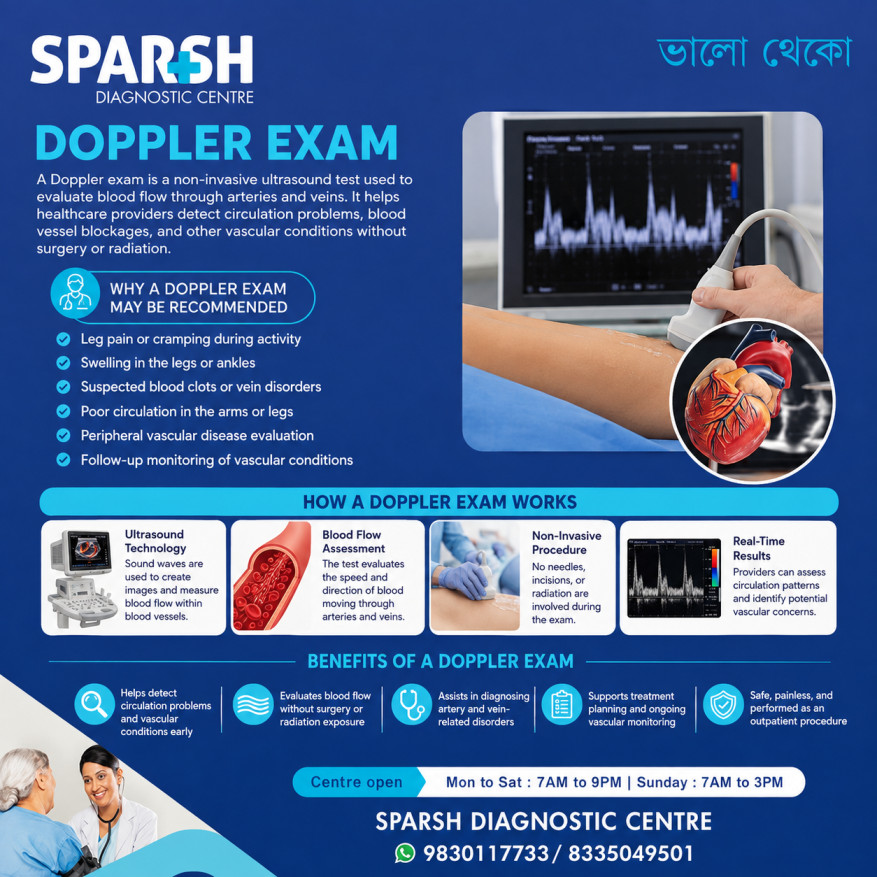
Can a Doppler ultrasound be used to diagnose blood clots?
Yes. A Doppler ultrasound can be used to help diagnose blood clots, including deep vein thrombosis (DVT). This is one of its most important applications.
When a clot forms in a deep vein, particularly in the leg, it can partially or completely interfere with normal blood flow. During a venous Doppler examination, the ultrasound specialist evaluates the veins, blood-flow pattern and whether the veins respond normally to gentle compression.
A clot may cause the affected vein to become difficult or impossible to compress and may alter the expected blood-flow pattern.
Doppler ultrasound is particularly valuable because it can provide this information without an injection or exposure to ionising radiation.
For suspected proximal DVT, ultrasound has generally shown high diagnostic accuracy. However, its ability to detect very small or distal clots can be lower, so the overall accuracy depends on the location of the clot, the technique used, the equipment and the experience of the person performing and interpreting the examination.
Can Doppler ultrasound detect blocked arteries?
Yes. Doppler ultrasound can help detect narrowed or blocked arteries.
When an artery becomes narrowed, blood has less space to pass through. The blood may accelerate as it moves through the narrowed section. Doppler measurements can identify abnormal flow velocities and patterns that suggest significant narrowing or obstruction.
A vascular Doppler examination may therefore help evaluate conditions such as:
- Peripheral artery disease (PAD)
- Arterial occlusion
- Narrowed arteries
- Reduced blood supply to the legs or arms
- Carotid artery narrowing
- Certain aneurysms
- Blood-vessel injuries
Doppler ultrasound can also be used to monitor blood flow after certain vascular procedures or treatments.
It is important to understand that the scan does not simply provide a yes-or-no answer in every case. The radiologist interprets the blood-flow velocities, waveforms, vessel appearance and other findings together to determine whether there is clinically significant narrowing.
What conditions can a leg Doppler detect?
A leg Doppler ultrasound can examine both the veins and arteries of the legs. The exact findings depend on whether a venous or arterial Doppler examination has been requested.
A venous Doppler may help detect:
- Deep vein thrombosis (DVT)
- Venous obstruction
- Chronic venous insufficiency
- Abnormal venous reflux
- Varicose-vein-related circulation problems
An arterial Doppler may help evaluate:
- Peripheral artery disease
- Narrowed arteries
- Blocked arteries
- Reduced arterial blood flow
- Arterial injuries
- Changes in blood flow after treatment
If you have symptoms such as one-sided swelling, persistent leg pain, unexplained cramping, cold feet or changes in skin colour, your doctor may recommend a Doppler examination to determine whether abnormal circulation could be contributing to the problem.
Why would a doctor order a Doppler ultrasound?
Doctors commonly order a Doppler ultrasound when they need to investigate symptoms or a medical condition that could be affecting blood flow.
Some common reasons include:
- Sudden or unexplained swelling in a leg
- Leg pain or tenderness
- Pain or cramping while walking
- Suspected DVT
- Coldness in the feet or hands
- Changes in skin colour
- Weak or reduced pulses
- Suspected peripheral artery disease
- Varicose veins or suspected venous insufficiency
- Monitoring a known vascular condition
- Checking blood flow after vascular treatment or surgery
- Evaluating certain blood-vessel injuries
Doppler ultrasound may also be used to examine blood flow in the neck, abdomen, heart and other parts of the body.
How does Doppler ultrasound help identify circulation problems?
The examination provides more than a simple picture of a blood vessel.
The ultrasound can show whether a vessel is open, while Doppler information helps demonstrate how blood is moving through it. The examination may reveal changes in:
- Blood-flow direction
- Blood-flow speed
- Flow pattern
- Vessel narrowing
- Vessel obstruction
- Venous reflux
- Areas where blood flow is reduced or absent
For example, if an artery is significantly narrowed, blood-flow velocity may increase at the site of narrowing and the downstream waveform may change. If a vein contains a significant clot, normal venous flow and compressibility may be affected.
This combination of structural and flow information makes vascular Doppler particularly useful for investigating circulation.
What happens during a Doppler scan?
A Doppler ultrasound is generally straightforward and does not require needles or injections.
You may be asked to remove clothing from the area being examined and lie or sit in a position that allows the sonographer to access the blood vessels.
A clear ultrasound gel is applied to the skin. The sonographer then moves the transducer over the area being examined.
You may hear soft whooshing sounds during the examination. These sounds are generated from the Doppler information and reflect blood movement.
For some leg examinations, blood-pressure cuffs may also be placed at different points on the limb to assess circulation.
The examination is generally painless, although you may experience mild pressure from the transducer, particularly when the sonographer is assessing veins.
How much time is taken for a Doppler scan?
A Doppler ultrasound commonly takes about 30 to 60 minutes, although the exact duration varies depending on the area being examined and the complexity of the study.
A straightforward examination of one region may take less time, while a more detailed study involving multiple blood vessels or both legs can take longer.
It is a good idea to allow some additional time for registration, changing and preparation rather than planning your appointment around the scan time alone.
How should I prepare for a Doppler ultrasound?
Preparation depends on the type of Doppler examination you are having.
For many leg or arm vascular Doppler examinations, little or no special preparation is required. You will usually need to wear or bring comfortable clothing that allows easy access to the area being examined.
However, some Doppler examinations may require fasting. Your doctor or diagnostic centre will tell you if this applies to your particular scan.
You may also be advised to avoid smoking or nicotine products for a period before certain examinations because nicotine can temporarily narrow blood vessels and potentially influence blood-flow measurements.
The safest approach is to follow the preparation instructions provided when your appointment is booked.
What are the risks of a Doppler ultrasound?
For most people, Doppler ultrasound is considered a safe, non-invasive and low-risk examination.
It does not use ionising radiation like X-rays or CT scans, and vascular Doppler examinations generally do not require an injected contrast dye.
You may feel some pressure from the ultrasound probe, but the procedure is usually painless.
As with other forms of ultrasound, the examination should be performed when medically appropriate and by trained professionals. The technology has an excellent safety record when used correctly.
What happens if a Doppler test is positive?
A “positive” Doppler result generally means that the examination has identified an abnormality, but the next step depends on exactly what was found.
For example, a positive venous Doppler may show evidence of DVT. An arterial Doppler may demonstrate narrowing, obstruction or reduced blood flow. A venous study may instead identify reflux or venous insufficiency.
The doctor will interpret the result alongside your symptoms, medical history and examination. Depending on the findings, you may need treatment, follow-up imaging, referral to a vascular specialist or additional tests.
If a Doppler identifies a suspected acute blood clot, particularly when symptoms are significant, medical assessment should not be delayed.
A Doppler report is therefore not simply about whether the result is “positive” or “negative”. The location, severity and nature of the abnormality are important.
How accurate is Doppler ultrasound?
Doppler ultrasound can be highly accurate when performed for the appropriate indication using good technique and interpreted by an experienced professional.
Its accuracy is particularly strong for many clinically significant proximal DVTs. Research has found high sensitivity and specificity for proximal DVT, although performance is generally lower for isolated distal DVT.
Accuracy can also be affected by factors such as:
- The location and size of the abnormality
- Patient movement
- Body habitus
- Vessel depth
- Ultrasound equipment
- Operator technique
- Doppler angle
- Machine settings
- The experience of the sonographer and interpreting radiologist
This is why a technically well-performed examination and appropriate clinical interpretation are both important.
What is the most common error associated with Doppler ultrasound?
One important source of error is incorrect Doppler angle or angle correction.
Doppler calculations of blood-flow velocity depend on the angle between the ultrasound beam and the direction of blood flow. As the angle approaches 90 degrees, the Doppler frequency shift becomes very small, making accurate velocity measurement difficult. For vascular velocity measurements, an angle of 60 degrees or less is generally preferred.
Other technical errors can result from inappropriate gain, incorrect velocity scale, poor sample-volume placement, patient movement or inadequate positioning.
This highlights why Doppler ultrasound is not simply a matter of placing a probe on the skin. Proper technique and knowledge of vascular ultrasound are essential for reliable results.
What is the most common artifact encountered in Doppler ultrasound?
Aliasing is one of the most commonly recognised artifacts in Doppler ultrasound.
It occurs when the blood-flow velocity being measured exceeds the system’s ability to display it correctly at the selected pulse repetition frequency or velocity scale. On colour Doppler, it can appear as an unexpected change between colour directions, while on pulsed-wave Doppler the waveform can appear to wrap around the baseline.
Aliasing does not automatically mean that something is wrong with the blood vessel. It can be a technical phenomenon caused by the selected Doppler settings, although it can also appear in areas of genuinely high blood-flow velocity, such as near a stenosis.
Other Doppler artifacts include blooming, motion artifacts, mirror-image artifacts and spectral broadening.
What do the results of a Doppler test for legs indicate?
The results of a leg Doppler provide information about how blood is moving through the examined veins or arteries.
A normal venous Doppler generally indicates that the examined veins are open and that there is no evidence of the abnormalities the scan was designed to detect, such as a significant clot.
An abnormal venous study may suggest DVT, venous obstruction or abnormal reflux.
For an arterial Doppler, the findings may indicate whether blood is flowing normally, whether an artery is narrowed or blocked, and whether there is evidence of reduced circulation.
The report may describe blood-flow velocities and waveforms, the condition of individual vessels and the location or severity of abnormalities.
Your doctor will interpret these findings in the context of your symptoms and other medical information.
How serious is poor circulation in the legs?
Poor circulation in the legs can range from a relatively mild problem to a serious vascular condition.
For example, chronic venous insufficiency can cause swelling, heaviness, skin changes and discomfort. Peripheral artery disease, on the other hand, occurs when arteries supplying the legs become narrowed or blocked. It may cause pain or cramping while walking and, in advanced cases, can contribute to wounds that heal poorly and tissue damage.
Significant arterial disease is also important because peripheral artery disease can be a marker of wider cardiovascular disease.
Symptoms should not be ignored, particularly if circulation problems are worsening. Sudden severe leg pain, marked swelling, a cold or pale limb, significant colour change or other acute symptoms require prompt medical assessment.
Why early evaluation matters
Blood clots and circulation disorders are often easier to manage when they are recognised promptly.
A Doppler ultrasound offers doctors a convenient way to investigate blood flow without radiation, surgery or invasive vascular procedures. It can help identify DVT, narrowed or blocked arteries, venous insufficiency and other vascular abnormalities.
If you have persistent leg swelling, unexplained pain, cramps while walking, cold feet or other symptoms suggesting a circulation problem, speak with your doctor about whether a vascular Doppler examination is appropriate.
Frequently asked questions
Can a Doppler ultrasound be used to diagnose blood clots?
Yes. Doppler and duplex ultrasound are commonly used to evaluate suspected DVT and other blood clots in the veins. The examination assesses the appearance and compressibility of the vein and evaluates blood flow.
Can Doppler ultrasound detect blocked arteries?
Yes. Doppler ultrasound can help identify narrowed or blocked arteries by assessing blood-flow patterns and velocities. It is commonly used to evaluate peripheral artery disease and arterial occlusion.
What are the risks of a Doppler ultrasound?
Doppler ultrasound is generally considered safe and non-invasive. It does not use ionising radiation and usually does not require contrast injections.
What happens if a Doppler test is positive?
A positive or abnormal result means that the scan has identified a finding that may require medical attention. The next step depends on whether the abnormality is a blood clot, arterial narrowing, blockage, venous reflux or another vascular problem.
How much time is taken for a Doppler scan?
Most Doppler examinations take around 30 to 60 minutes, although the duration depends on the area being examined and the complexity of the study.
What is the most common error associated with Doppler ultrasound?
Incorrect Doppler angle or angle correction is an important source of velocity-measurement error. Other errors can result from inappropriate machine settings, patient movement and poor sample positioning.
What conditions can a leg Doppler detect?
A leg Doppler can help detect DVT, venous insufficiency, venous obstruction, peripheral artery disease, arterial narrowing, arterial blockage and other abnormalities affecting blood flow in the legs.
How should I prepare for a Doppler ultrasound?
Many leg Doppler examinations require little or no special preparation. Depending on the type of examination, you may be asked to fast or avoid nicotine beforehand. Always follow the instructions provided by your doctor or diagnostic centre.
What is the most common artifact encountered in Doppler ultrasound?
Aliasing is a common Doppler artifact that occurs when measured flow velocity exceeds the selected Doppler scale or pulse repetition frequency. It can affect both colour and pulsed-wave Doppler.
Why would a doctor order a Doppler ultrasound?
A doctor may order one to investigate suspected blood clots, poor circulation, arterial narrowing or blockage, venous insufficiency, vascular injury or to monitor an existing vascular condition.
How accurate is Doppler ultrasound?
Doppler ultrasound can be highly accurate, particularly for many proximal DVTs, but accuracy varies depending on the condition, location of the abnormality, technique and operator experience. Distal DVT can be more difficult to detect.
What do the results of a Doppler test for legs indicate?
The results indicate how blood is flowing through the examined arteries or veins and whether there is evidence of abnormalities such as a clot, narrowing, blockage or abnormal venous flow.
How serious is poor circulation in legs?
The seriousness depends on the underlying cause and severity. Mild venous circulation problems may cause swelling and discomfort, while significant arterial disease can threaten tissue health and may indicate a higher risk of cardiovascular disease. New or severe symptoms should be assessed promptly.
Conclusion
Doppler ultrasound has become an important tool for investigating blood clots and circulation problems because it provides valuable information about blood flow without the need for radiation or invasive procedures.
From suspected DVT and leg swelling to peripheral artery disease and blocked blood vessels, a Doppler examination can help doctors understand what is happening inside the circulatory system. It can also be useful for monitoring known vascular conditions and assessing treatment.
If your doctor has recommended a Doppler ultrasound, there is usually no reason to be anxious about the procedure. It is generally painless, non-invasive and straightforward. Most importantly, it can provide information that helps your doctor decide whether further evaluation or treatment is needed.
For anyone experiencing unexplained leg swelling, persistent pain, walking-related cramps, changes in skin colour, cold feet or other symptoms of poor circulation, timely medical evaluation can make a meaningful difference.
Book Your Doppler Ultrasound Today at Sparsh Diagnostic Centre
At Sparsh Diagnostic Centre, we use state-of-the-art Doppler technology to deliver fast, accurate, and reliable diagnostics. Our expert radiologists ensure you receive the best possible care for your vascular health.
📞 Call: 9830117733 / 8335049501
🌐 Visit: www.sparshdiagnostica.com
#BhaloTheko
Disclaimer:
No content on this site, regardless of date, should ever be used as a substitute for direct medical advice from your doctor or other qualified clinician.


[…] Doppler Ultrasound: May detect flow abnormalities in the splenic vein but less sensitive in obese or gassy patients. […]
[…] The most common and non-invasive test for diagnosing DVT is a Doppler ultrasound. This test uses sound waves to visualize blood flow in the veins and detect […]
[…] Ultrasound with Doppler: Assesses portal vein diameter and flow […]
[…] Ultrasound with Doppler – First-line imaging to assess blood flow in the portal vein. […]
[…] Doppler Ultrasound in […]
[…] 4. Doppler Ultrasound […]
[…] 4. Ultrasound with Doppler […]
[…] Doppler ultrasound: A Doppler scan uses sound waves to assess blood flow in the placenta, umbilical cord, and fetus. It is often used to check the health and well-being of the fetus during the second and third trimesters. […]
[…] 3. Doppler Ultrasound […]