
Medical imaging has revolutionized the way doctors diagnose and treat diseases. Among the most advanced imaging tools available today is the CT Scan (Computed Tomography Scan). It offers detailed cross-sectional images of the body, enabling doctors to detect abnormalities, plan surgeries, and monitor treatments with precision.
If you’ve been recommended a CT scan, you might have questions about what it is, how it works, and what to expect. This guide will walk you through everything you need to know about CT scans, including their uses, benefits, risks, and preparation.
What is a CT Scan?
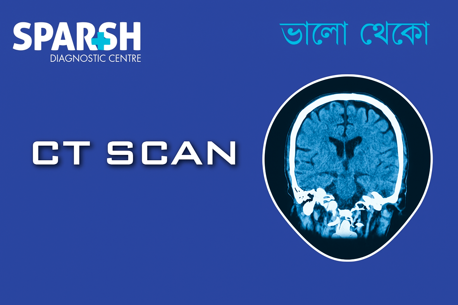
A CT scan (also known as CAT scan or Computed Tomography scan) is a medical imaging technique that uses X-rays and computer processing to create detailed cross-sectional images of the body. Unlike a regular X-ray, which shows only a flat image, a CT scan provides multiple layered images that can be reconstructed into 3D views.
This allows doctors to see bones, organs, blood vessels, and soft tissues with much greater clarity.
How Does a CT Scan Work?
The CT scanner is a large machine shaped like a doughnut. The patient lies on a motorized table that slides into the scanner. The machine rotates around the body, emitting X-rays from different angles. Detectors capture the X-rays, and a computer processes the data to produce detailed images.
Sometimes, a contrast dye is used to enhance visibility of certain structures such as blood vessels, intestines, or tumors.
Types of CT Scans
Different CT scans are performed depending on the medical condition:
Head CT Scan – To detect strokes, tumors, bleeding, or skull fractures.
Chest CT Scan – For lung diseases, pulmonary embolism, heart abnormalities, or infections.
Abdominal and Pelvic CT Scan – To diagnose kidney stones, appendicitis, cancers, or abdominal pain.
Cardiac CT Scan – Used to visualize heart structures, blockages, or congenital defects.
CT Angiography – Special scan to evaluate blood vessels for clots, aneurysms, or blockages.
Spine CT Scan – For herniated discs, spinal stenosis, or fractures.
Whole Body CT Scan – Used for trauma cases or preventive health screenings.
Why is a CT Scan Done?
Doctors recommend CT scans for various reasons, including:
Detecting internal injuries or bleeding after accidents.
Diagnosing tumors, infections, or chronic diseases.
Guiding biopsies and minimally invasive procedures.
Planning radiation therapy for cancer patients.
Monitoring treatment progress.
Detecting bone fractures and joint problems.
Evaluating blood clots and vascular conditions.
Benefits of a CT Scan
CT scans offer numerous advantages over other imaging techniques:
High accuracy – Provides detailed and precise images.
Quick and painless – Usually completed within 10–30 minutes.
Non-invasive – No surgical instruments are required.
Comprehensive view – Detects abnormalities in multiple body parts simultaneously.
Guidance tool – Helps doctors plan surgeries and treatments effectively.
Risks of a CT Scan
Like all medical procedures, CT scans come with certain risks:
Radiation exposure – Although the dose is relatively low, repeated scans can increase risk.
Allergic reaction to contrast dye – Some patients may experience mild reactions like rash or nausea.
Kidney issues – Patients with kidney disease should consult their doctor before contrast scans.
Pregnancy concerns – CT scans are usually avoided during pregnancy unless absolutely necessary.
However, the benefits of early diagnosis usually outweigh the risks.
How to Prepare for a CT Scan
Preparation depends on the type of scan:
With contrast dye – You may need to avoid food and drinks for 4–6 hours before the scan.
Without contrast – Minimal preparation is required.
Wear loose-fitting clothes; you may be asked to change into a hospital gown.
Remove metal objects like jewelry, glasses, and belts.
Inform the radiologist about pregnancy, kidney disease, diabetes, or allergies.
What Happens During a CT Scan?
You will lie on a motorized table that slides into the CT scanner.
If contrast is needed, it may be given orally, intravenously, or rectally.
The scanner rotates around your body, taking images.
You may be asked to hold your breath briefly to avoid motion blur.
The entire process usually takes 10–30 minutes.
What to Expect After a CT Scan
You can usually resume normal activities immediately.
If contrast dye was used, you may be advised to drink plenty of fluids to flush it out.
Your results are analyzed by a radiologist and shared with your doctor.
Cost of a CT Scan in India
The cost of a CT scan depends on:
Type of CT scan (head, chest, abdomen, angiography, etc.).
Whether contrast dye is used.
City and healthcare facility.
On average, CT scan prices in India range between ₹2,000 to ₹10,000.
Frequently Asked Questions (FAQ)
1. Is a it painful?
No, CT scans are painless. You may only feel slight discomfort while lying still or from the injection of contrast dye.
2. How long does a it take?
Most CT scans are completed within 10–30 minutes.
3. Can I eat or drink before the scan?
If your scan requires contrast dye, you may be asked to avoid eating or drinking for 4–6 hours before. Otherwise, you can eat normally.
4. Are CT scans safe?
Yes, CT scans are generally safe. While they involve low radiation exposure, the benefits outweigh the risks in most cases.
5. Can I get a CT scan during pregnancy?
CT scans are usually avoided during pregnancy unless urgently needed. An ultrasound or MRI is preferred.
6. How soon will I get my CT scan results?
At most diagnostic centres, including Sparsh Diagnostic Centre, results are available within a few hours to 1–2 days.
7. What is the difference between a CT scan and an MRI?
CT scans use X-rays and are faster, while MRIs use magnetic fields and provide more detail of soft tissues.
A CT scan is a highly effective diagnostic tool that provides doctors with detailed images of the body. Whether it’s detecting a disease, guiding treatment, or monitoring recovery, CT scans play a vital role in modern medicine.
If your doctor has recommended a CT scan, there’s no need to worry — the procedure is quick, safe, and reliable.
#BhaloTheko
Disclaimer:
No content on this site, regardless of date, should ever be used as a substitute for direct medical advice from your doctor or other qualified clinician.

![]()


[…] CT scans (trauma) […]
[…] CT Pulmonary Angiography: […]
[…] CT scan – assesses spread to lymph nodes and organs […]
[…] and CT Scans – Detect brain or spinal cord […]
[…] CT scan if MRI is unavailable […]
[…] CT Scan of the Chest: Provides detailed visualization of the abscess, surrounding tissues, and any complications. […]
[…] CT scan or ultrasound (for abscesses or organ involvement) […]
[…] CT scans or MRI may be used to evaluate structural abnormalities or pelvic conditions. […]
[…] X-ray or CT scan – to rule out lung […]
[…] CT scan in selected cases […]
[…] X-ray or CT scan: If infection has […]
[…] Studies: Imaging techniques like chest X-rays, CT scans, and ultrasounds can help detect the source of infection, such as pneumonia or an abdominal […]
[…] CT scan or MRI to identify abscesses or organ involvement […]
[…] 4. CT Scan […]
[…] CT or MRI (in rare […]
[…] CT/MRI brain – Rule out stroke or tumor […]
[…] CT scan – Provides detailed imaging for complex cases. […]
[…] CT scan […]
[…] or CT scans may be used to rule out herniated discs or […]
[…] 4. Imaging Tests (X-ray, CT Scan) […]
[…] CT scan of chest […]
[…] CT scans […]
[…] CT Scan […]
[…] tests such as CT or MRI (in some […]
[…] CT Scan: A CT scan of the abdomen and pelvis may be ordered to assess the extent of inflammation and rule out complications such as abscesses or perforation. […]
[…] CT scan: Helps detect complications like perforation. […]
[…] CT or MRI Scans: In some cases, doctors may use a CT or MRI scan to provide more detailed images of the veins and detect clots that are not easily visible on an ultrasound. […]
[…] 4. What if my Doppler test is negative, but I still have symptoms?Your doctor may repeat the test after a few days or order additional tests like a D-dimer or CT scan. […]
[…] CT scan or MRI – detailed liver architecture […]
[…] 3. MRI or CT Scan […]
[…] Ultrasound/CT scan – For suspected amoebic liver abscess […]
[…] CT or MR […]
[…] CT Scan […]
[…] X-ray or CT scan can reveal emphysematous changes or airway wall […]
[…] CT or MRI Brain – to rule out structural brain damage or stroke. […]
[…] or CT Scan: To check for stroke, tumor, or inner ear […]
[…] X-ray or CT Scan: Detects structural lung […]
[…] CT Scan with Myelogram: Highlights nerve root involvement using contrast dye. […]
[…] Tests: MRI or CT scans may be ordered to rule out other causes of sciatic nerve […]
[…] 3. MRI or CT Scan […]
[…] Chest X-ray or CT scan […]
[…] CT Scan: Evaluates deep tissue involvement. […]
[…] CT Scan: Provides cross-sectional views for detailed bone and tissue analysis. […]
[…] CT or MRI of the liver […]
[…] CT or MRI Scan:Imaging tests to visualize inflammation, abscesses, or fistulas. […]
[…] CT Scan: Offers cross-sectional images of the spine to reveal nerve impingements or abnormalities. […]
[…] 3. CT Scan (Computed Tomography) […]
[…] CT Scan: Provides detailed imaging for conditions like pulmonary embolism or interstitial lung disease. […]
[…] 3. CT Scan of the Chest […]
[…] CT Scan (Computed Tomography): Quickly detects bleeding, swelling, or structural abnormalities. […]
[…] or CT Scan: Detects brain swelling, bleeding, or structural […]
[…] CT Scan: […]
[…] 3. High-Resolution CT (HRCT) Scan […]
[…] 5. Chest X-Ray or CT Scan […]
[…] X-rays or CT scans can detect pneumonia, embolism, or other lung […]
[…] CT Scan or MRI:Used when more detailed imaging is needed, especially for pulmonary embolism or cardiac […]
[…] CT or MRI scans: To detect internal bleeding or blockages […]
[…] tests like X-rays, MRI, or CT scans to assess the […]
[…] CT Scan (Computed Tomography): Helps detect skull base fractures and fluid accumulation. […]
[…] Tests – CT, MRI, or PET scans help determine the extent of […]
[…] 2. CT Scan (Computed Tomography) […]
[…] Tests: Imaging tests such as a CT scan, MRI, or PET scan may be used to determine the extent of the cancer and whether it has spread to […]
[…] Tests (MRI/CT): To check for neurological or structural […]
[…] CT Scan: A computed tomography (CT) scan may provide detailed images of the lungs and chest to diagnose conditions like pulmonary embolism or lung disease. […]