


Pregnancy brings a long list of appointments, blood tests and scans, and pregnancy ultrasonography tests are among the most important. An ultrasound uses high-frequency sound waves to create images of the developing baby, uterus, placenta and surrounding structures. Unlike X-rays, ultrasound does not use ionising radiation.
But there is no single ultrasound schedule that applies to every pregnancy. Some women may have only the routine scans, while others may need additional scans because of their medical history, symptoms, multiple pregnancy, concerns about fetal growth or findings on an earlier examination.
In many pregnancy-care systems, two major scans are routinely offered: a first-trimester dating scan around 11–14 weeks and a detailed anomaly scan around 18–21 weeks. Additional examinations may be recommended when clinically necessary.
So, what exactly happens during these scans? When should you have them? And what does it mean if an early ultrasound does not show a heartbeat?
Let’s take a closer look.
What is pregnancy ultrasonography?
Pregnancy ultrasonography, commonly called a pregnancy ultrasound, obstetric ultrasound or pregnancy sonography, is an imaging test used to assess pregnancy and fetal development.
Depending on the stage of pregnancy, an ultrasound can help assess:
- Location of the pregnancy
- Gestational age
- Fetal heartbeat or cardiac activity
- Number of babies
- Fetal growth
- Baby’s anatomy
- Placenta
- Amniotic fluid
- Cervix and uterus
- Blood flow through the umbilical cord and other vessels
- Certain structural abnormalities
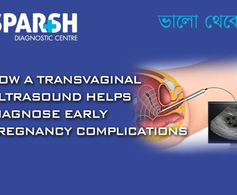
Ultrasound may be performed through the abdomen or, particularly in early pregnancy, through the vagina using a transvaginal probe.
How many sonography tests are there in pregnancy?
There is no fixed number of sonography tests that every pregnant woman must have.
A low-risk pregnancy may have two or more routine scans, while a pregnancy requiring closer monitoring may involve several additional examinations. ACOG notes that at least one standard ultrasound is generally performed during pregnancy, usually around 18–22 weeks in the US, while a first-trimester scan may also be performed.
In Indian practice, doctors may recommend a more detailed schedule depending on the pregnancy and the patient’s individual needs.
Common pregnancy ultrasound examinations include:
- Early pregnancy or viability scan
- Dating scan
- Nuchal translucency (NT) scan
- First-trimester screening ultrasound
- Anomaly scan
- Fetal echocardiography, when indicated
- Growth or pregnancy-wellbeing scan
- Obstetric Doppler
- Follow-up scans for high-risk pregnancies
These are not nine mandatory scans. Several names can refer to different components of the same examination.

What are the 5 scans in pregnancy?
When people refer to the “five scans in pregnancy”, they usually mean a commonly discussed sequence rather than a universal medical requirement.
Five important scans may include:
1. Early pregnancy or dating scan
Usually performed in early pregnancy when clinically indicated, it can confirm the location of the pregnancy, estimate gestational age and assess early development.
2. NT or first-trimester scan
Usually performed around 11–14 weeks. It may include measurement of nuchal translucency and other markers as part of first-trimester screening.
3. Anomaly scan
Usually performed around 18–22 weeks. It provides a detailed assessment of the baby’s anatomy and development.
4. Growth scan
A later-pregnancy scan may assess fetal growth, presentation, amniotic fluid and other parameters when recommended.
5. Obstetric Doppler
Doppler ultrasound evaluates blood flow through selected maternal and fetal blood vessels. It is particularly useful when there is concern about fetal growth, placental function or other pregnancy complications.
The exact timing should always be determined by the obstetrician or fetal-medicine specialist rather than following a generic internet schedule.
Pregnancy ultrasound timeline by trimester
First trimester: early pregnancy to 13 weeks
An early scan may be recommended to confirm an intrauterine pregnancy, estimate gestational age, assess development and, when appropriate, detect cardiac activity.
The dating scan is generally performed around 10–14 weeks in many care pathways. It helps determine how far the pregnancy has progressed and may be combined with screening.
Second trimester: 14–27 weeks
This is when the detailed anomaly scan becomes particularly important.
The 20-week scan, also called the mid-pregnancy or anomaly scan, examines the baby’s anatomy and looks for signs of several structural conditions. It also assesses growth, placenta and certain aspects of blood flow.
A fetal echocardiography may also be recommended when there is an increased risk of congenital heart disease or a concern identified on another examination.
Third trimester: 28 weeks onward
Later pregnancy scans are generally performed when clinically indicated. They may include:
- Growth scan
- Amniotic fluid assessment
- Placental assessment
- Fetal position
- Biophysical assessment
- Obstetric Doppler
- Follow-up of previously identified concerns
A scan around seven months is therefore often described as a growth scan or pregnancy-wellbeing scan, sometimes combined with Doppler depending on the clinical situation. There isn’t one universally defined “7-month scan.”
How is a 7-week ultrasound done?
At around seven weeks, an ultrasound may be performed transvaginally, particularly if the pregnancy is very early or abdominal imaging does not provide sufficient detail.
During a transvaginal scan, a specially designed covered ultrasound probe is gently inserted into the vagina. The examination allows the sonographer or doctor to obtain closer images of the uterus and early pregnancy.
Depending on the actual gestational age, the scan may show a gestational sac, yolk sac, embryo and cardiac activity.
The exact appearance varies considerably depending on when ovulation occurred, implantation timing, menstrual-cycle length and the quality of the images.
Is an empty sac at 7 weeks normal?
An empty gestational sac at a presumed seven weeks does not automatically mean a miscarriage.
Pregnancy dates can be wrong, particularly when ovulation occurred later than expected. Sometimes a repeat ultrasound is necessary before a definite diagnosis can be made.
ACOG specifically notes that a single ultrasound may not always be sufficient to confirm early pregnancy loss, and conservative ultrasound criteria are used to avoid incorrectly diagnosing a potentially viable pregnancy.
If an ultrasound shows an empty sac, your doctor may recommend a repeat examination after an appropriate interval, sometimes alongside serial hCG testing.
What if there’s no heartbeat at 6 weeks?
Not seeing cardiac activity at six weeks does not automatically mean that the pregnancy has been lost.
The timing of ovulation and implantation can vary, and even a difference of several days can significantly change what is visible on an early scan.
ACOG notes that cardiac activity may simply be too early to detect, although in some cases its absence can indicate that the embryo has stopped developing. Further ultrasound examinations and, in some cases, hCG testing may be needed.
Is no heartbeat a miscarriage?
Not necessarily.
A diagnosis of miscarriage should not be made simply because cardiac activity is not seen at six weeks. The ultrasound findings, measurements, dates and clinical circumstances all matter.
If the scan is inconclusive, your doctor may recommend waiting and repeating the ultrasound rather than making an immediate diagnosis.
How do I know my baby is okay at 6 weeks?
At six weeks, an ultrasound can provide useful information, but it cannot guarantee that everything will remain normal throughout pregnancy.
Depending on the actual gestational age, an ultrasound may show:
- Gestational sac
- Yolk sac
- Embryo
- Crown-rump length (CRL)
- Cardiac activity
- Location of the pregnancy
A reassuring scan is encouraging, but pregnancy continues to develop rapidly, which is why later scans remain important.
How accurate is an 8-week ultrasound?
An ultrasound around eight weeks can be very useful for dating an early pregnancy, particularly when the crown-rump length can be measured clearly.
However, no ultrasound is 100% accurate. Dating becomes an estimate based on measurements, and differences can occur because of variations in ovulation, implantation, fetal position and measurement technique.
First-trimester crown-rump-length measurement is widely used for estimating gestational age because it provides a useful early measurement of fetal size.
What are good signs at 8 weeks pregnant?
Reassuring ultrasound findings may include:
- A pregnancy located inside the uterus
- Appropriate development for the estimated gestational age
- Visible embryo
- Appropriate crown-rump length
- Cardiac activity
- Expected development of early pregnancy structures
Importantly, symptoms such as nausea or breast tenderness are not reliable tests of fetal wellbeing. The ultrasound and clinical assessment provide more useful information.
Why do doctors sometimes wait until 8 weeks?
Doctors may recommend waiting until around seven or eight weeks because the pregnancy is easier to evaluate than it would be at four or five weeks.
Waiting can make it more likely that the scan will show the embryo and cardiac activity clearly, reducing uncertainty and the need for an immediate repeat scan.
That said, an earlier ultrasound can be appropriate when there is bleeding, pain, previous pregnancy complications, fertility treatment, suspected ectopic pregnancy or another clinical reason.
Can a 4-week pregnancy be seen on ultrasound?
Sometimes, but often not much can be seen at four weeks.
At this stage, the pregnancy may be too early for an ultrasound to provide meaningful information. This is one reason an ultrasound performed too early can create unnecessary anxiety.
The doctor may instead recommend waiting until the pregnancy is further developed, depending on the clinical situation.
Is ultrasound 100% correct?
No.
Ultrasound is an extremely useful diagnostic tool, but it is not 100% accurate.
The quality and accuracy of an examination can be affected by:
- Gestational age
- Baby’s position
- Maternal body habitus
- Amount of amniotic fluid
- Uterine position
- Movement
- Image quality
- Equipment
- Experience of the operator
- The type of abnormality being assessed
Even the detailed anomaly scan cannot detect every possible fetal condition. The NHS notes that some conditions are easier to see than others.
Can ultrasound give wrong results?
Ultrasound can occasionally produce an incorrect, uncertain or incomplete result.
This does not necessarily mean the technology “failed.” Sometimes the scan simply cannot provide enough information at that moment.
For example, an early scan may suggest that a pregnancy is younger than expected. A baby’s position may also prevent the sonographer from obtaining certain views during an anatomy scan.
If an image is unclear or a finding is uncertain, a repeat ultrasound or a more specialized examination may be recommended.
Which gender is easier to see on ultrasound?
There is no reliable rule that one fetal sex is “easier” to see than the other.
Visibility depends mainly on:
- Gestational age
- Baby’s position
- Image quality
- The viewing angle
- Experience of the examiner
In India, prenatal determination and communication of fetal sex are prohibited under the PCPNDT Act, so ultrasound examinations should not be used to determine or disclose fetal sex.
The purpose of pregnancy ultrasound is to assess the health and development of the pregnancy, not to determine fetal sex.
How do I read my pregnancy ultrasound report?
Ultrasound reports can look confusing because they contain abbreviations and measurements. Some common terms include:

Can I eat before an ultrasound?
Usually, yes, unless your healthcare provider or diagnostic centre gives you different instructions.
Pregnancy ultrasound generally does not require fasting. However, preparation can vary according to the type of scan.
If you’re having another test at the same appointment, such as a blood test requiring fasting, follow those specific instructions.
Can I drink water before an ultrasound?
It depends on the type of examination.
For some early abdominal or pelvic ultrasound examinations, a full bladder may improve visualization. Some centres therefore ask patients to drink water before the scan.
For a transvaginal ultrasound, a full bladder is generally not required.
The safest approach is to follow the preparation instructions given by the diagnostic centre. For example, NHS guidance notes that a full bladder may be requested for some dating scans to improve the image.
What is the most common ultrasound mistake?
One of the most common problems is interpreting the scan without considering the pregnancy dates and clinical context.
For example, someone may believe they are exactly seven weeks pregnant based on the last menstrual period, when they actually ovulated several days later. An ultrasound can then appear “behind” even though the pregnancy may simply be younger than expected.
Another common mistake is assuming that ultrasound can detect every abnormality or provide a guaranteed prediction of fetal health.
The best interpretation combines the ultrasound findings with menstrual history, previous scans, blood tests and the advice of the treating doctor.
How long do ultrasound results take?
The sonographer or radiologist may be able to discuss some findings during or immediately after the examination. In many settings, the formal report is also available the same day or shortly afterwards.
The exact turnaround time depends on the diagnostic centre, type of examination and whether specialist review is required.
For detailed fetal medicine or complicated cases, additional review may take longer.
Which pregnancy scan is the most important?
Rather than asking which single scan is “most important”, it is better to understand that different scans answer different questions.
The early scan helps establish the pregnancy and its timing. The first-trimester scan can contribute to early screening. The anomaly scan provides a detailed assessment of fetal anatomy. Later growth and Doppler scans can monitor pregnancies where additional surveillance is needed.
For example, the standard mid-pregnancy ultrasound is usually performed around 18–22 weeks and is designed to evaluate fetal anatomy in much greater detail than an early scan.
Which is the toughest month of pregnancy?
There is no medically defined “toughest month.”
Many women find the first trimester physically challenging because of nausea, vomiting, fatigue and hormonal changes. Others find the third trimester more difficult because of increasing abdominal size, back discomfort, sleep difficulties and frequent urination.
Every pregnancy is different, and even the same woman may experience different symptoms in different pregnancies.
What should you do before a pregnancy ultrasound?
A few simple steps can make the appointment easier:
- Follow the centre’s instructions about drinking water.
- Ask whether you need a full bladder.
- Carry previous ultrasound reports.
- Carry relevant blood-test reports.
- Know the first day of your last menstrual period if possible.
- Take your pregnancy-related medical records.
- Wear comfortable clothing.
- Tell the doctor about bleeding, pain or other symptoms.
- Don’t hesitate to ask questions about anything written in the report.
Most importantly, don’t compare your ultrasound measurements directly with someone else’s pregnancy. A measurement that is appropriate for one gestational age may be completely different at another stage.
When should you contact your doctor?
Pregnancy ultrasound is only one part of antenatal care. If you experience significant vaginal bleeding, severe abdominal or pelvic pain, dizziness, fainting, fluid leakage or any other concerning symptom, contact your obstetrician or seek appropriate medical care rather than waiting for a routine scan.
Similarly, if an early ultrasound shows no cardiac activity, an empty sac or uncertain findings, speak with your doctor before assuming that the pregnancy has been lost. A repeat scan may be necessary to establish what is happening.
Frequently Asked Questions About Pregnancy Ultrasonography Tests
How many sonography tests are in pregnancy?
There is no fixed number. Low-risk pregnancies may need only a few routine scans, while high-risk pregnancies may require additional growth, Doppler or specialized examinations.
What are the 5 scans in pregnancy?
A commonly discussed sequence includes an early/dating scan, NT scan, anomaly scan, growth scan and obstetric Doppler. However, these are not five compulsory scans for every pregnancy.
What is a 7-month scan called?
It is commonly called a growth scan, pregnancy-wellbeing scan or third-trimester ultrasound. Doppler may be added when clinically indicated.
How is a 7-week ultrasound done?
It may be performed transvaginally to obtain clearer images of the early pregnancy. A transabdominal examination may also be used depending on the circumstances.
Is an empty sac at 7 weeks normal?
It can be too early to make a definite diagnosis, particularly when dates are uncertain. A repeat ultrasound may be required.
When is the best time for the first ultrasound?
The best timing depends on the reason for the scan. For routine dating and first-trimester assessment, many care pathways use approximately 10–14 weeks, while an earlier scan may be indicated for specific clinical reasons.
What if there’s no heartbeat at 6 weeks?
It may simply be too early. The doctor may recommend a repeat scan rather than diagnosing miscarriage from this finding alone.
Is no heartbeat a miscarriage?
Not necessarily. Diagnosis requires appropriate ultrasound findings and measurements, and sometimes follow-up scanning.
How accurate is an 8-week ultrasound?
It can provide a very useful estimate of gestational age, especially when crown-rump length is measured, but it is not 100% accurate.
What are good signs at 8 weeks pregnant?
A visible embryo, appropriate growth and cardiac activity are reassuring ultrasound findings. Your doctor will interpret them in relation to gestational age.
Why do doctors wait until 8 weeks?
Waiting can make the pregnancy easier to visualize and may reduce uncertainty that can occur with very early scanning.
Is ultrasound 100% correct?
No. Ultrasound is highly useful but cannot detect every condition or guarantee a completely normal pregnancy.
Can ultrasound give wrong results?
Yes. Dates, fetal position, image quality and operator or technical factors can sometimes result in uncertain or misleading findings.
Which gender is easier to see on ultrasound?
Neither is inherently easier to see. However, fetal sex determination and disclosure through prenatal diagnostic techniques are prohibited in India under the PCPNDT Act.
How do I read my pregnancy ultrasound?
Look for terms such as CRL, GA, FHR/FCA, BPD, HC, AC, FL, EFW, AFI, placenta and Doppler. Ask your doctor to interpret the measurements as a group rather than relying on individual numbers.
Can I eat before an ultrasound?
Usually yes, unless your diagnostic centre specifically instructs you to fast.
Can I drink water before an ultrasound?
For some scans, particularly certain abdominal examinations, you may be asked to have a full bladder. Follow the instructions provided for your specific scan.
What is the most common ultrasound mistake?
A common mistake is assuming that one measurement or an early scan gives the complete picture. Ultrasound findings must be interpreted according to gestational age and the clinical situation.
How long do ultrasound results take?
Some results can be discussed immediately, while the formal report may be issued the same day or later depending on the centre and complexity of the examination.
Final thoughts
Pregnancy ultrasonography tests are an important part of modern antenatal care. From confirming an early pregnancy and estimating gestational age to examining fetal anatomy and monitoring growth and blood flow, different ultrasound examinations serve different purposes.
One important point to remember is that more scans do not necessarily mean a healthier pregnancy, and fewer scans do not necessarily mean something is wrong. The appropriate schedule depends on the individual pregnancy.
If an early scan doesn’t show a heartbeat, the gestational sac appears empty, or a measurement doesn’t match your expected dates, don’t jump to conclusions. Early pregnancy develops rapidly, and repeat imaging is sometimes necessary before a diagnosis can be made.
Likewise, a normal ultrasound is reassuring but cannot identify every possible pregnancy complication. Regular antenatal appointments and following your obstetrician’s recommendations remain essential.
For a pregnancy ultrasound, choose a diagnostic centre where obstetric imaging is performed by appropriately qualified professionals and where the findings can be properly interpreted in the context of your pregnancy.
Medical note: Pregnancy scan schedules vary by country, doctor and individual pregnancy. This article is for educational purposes and should not replace advice from your obstetrician or radiologist.
If you still have questions or need to consult a Gynecologist, do not hesitate to contact Sparsh Diagnostic Centre on 9830117733.
#BhaloTheko
Disclaimer:
No content on this site, regardless of date, should ever be used as a substitute for direct medical advice from your doctor or other qualified clinician.



[…] Ultrasound: An ultrasound may be ordered to detect the presence of a fetus, although as mentioned earlier, the baby’s position may sometimes make it difficult to see. […]
[…] Ultrasound: Identifies structural abnormalities in the kidneys. […]
[…] 5. Obstetric Ultrasound […]
[…] Ultrasound for Growth Monitoring […]
[…] Insight: Blood tests and early ultrasounds can confirm pregnancy and estimate gestational […]
[…] What is an Ultrasound Scan in Pregnancy? […]
[…] Ultrasound – Identifies fetal abnormalities like heart defects and growth restrictions. […]
[…] Ultrasound: Evaluates kidney size and structure. […]
[…] Studies: In severe or recurrent cases, imaging tests like ultrasound, CT scans, or MRI may be used to detect abnormalities in the urinary […]
[…] Translucency (NT) scan is one of the most crucial tests conducted during the first trimester. This non-invasive ultrasound test is instrumental in assessing the risk of certain chromosomal abnormalities, such as Down syndrome, […]
[…] Ultrasound: The most common and effective method for detecting gallstones. […]
[…] Tests: X-rays, ultrasounds, or CT scans to detect abnormalities in the […]
[…] echocardiography is a specialized ultrasound technique used to evaluate the heart of a developing fetus. This non-invasive procedure provides crucial […]
[…] Ultrasound: Uses sound waves to produce images of the inside of the breast. This is particularly useful for distinguishing between solid masses (like fibroadenomas) and cysts. […]
[…] triiodothyronine (T3) can help confirm the diagnosis. Additional imaging studies, such as thyroid ultrasound or radioactive iodine uptake scans, may be ordered to assess the structure and function of the […]