

Obstructive shock is a life-threatening medical emergency that occurs when blood flow is mechanically blocked, preventing the heart from pumping effectively. Unlike other forms of shock such as hypovolemic or cardiogenic shock, obstructive shock is caused by a physical obstruction that restricts blood circulation, leading to reduced cardiac output and inadequate oxygen delivery to tissues.
Without rapid diagnosis and treatment, obstructive shock can quickly lead to organ failure and death. Understanding its causes, symptoms, and management strategies is critical for both healthcare professionals and patients.
In this comprehensive guide, we’ll explore everything you need to know about obstructive shock, including its pathophysiology, major causes, clinical features, diagnostic approach, treatment options, and frequently asked questions.
What Is Obstructive Shock?
Obstructive shock is a type of circulatory shock caused by mechanical obstruction of blood flow in the heart or great vessels. This obstruction prevents adequate cardiac filling or output, leading to decreased tissue perfusion.
Key Characteristics:
Reduced venous return to the heart
Increased afterload
Decreased cardiac output
Inadequate oxygen delivery to vital organs
Obstructive shock is considered a medical emergency requiring immediate intervention to remove the obstruction.

Pathophysiology of Obstructive Shock
To understand obstructive shock, it’s important to review how normal circulation works.
Under normal conditions:
Blood returns to the heart via the superior and inferior vena cava.
The heart pumps blood into the pulmonary circulation.
Oxygenated blood returns to the left heart and is distributed systemically.
In obstructive shock, a blockage interferes with this process. The obstruction may occur:
In the pulmonary vasculature (e.g., pulmonary embolism)
Around the heart (e.g., cardiac tamponade)
In the thoracic cavity (e.g., tension pneumothorax)
This leads to:
Increased pressure inside the chest
Reduced venous return
Impaired ventricular filling
Decreased stroke volume
Hypotension and tissue hypoxia
If untreated, this cascade can result in multi-organ failure.
Types and Causes of Obstructive Shock
Several conditions can cause obstructive shock. The most common include:
1. Pulmonary Embolism (PE)
A massive pulmonary embolism occurs when a blood clot blocks the pulmonary arteries, preventing blood flow from the right side of the heart to the lungs.
How It Causes Shock:
Increased pulmonary vascular resistance
Right ventricular strain and failure
Reduced left ventricular filling
Decreased cardiac output
Symptoms may include sudden shortness of breath, chest pain, and hypotension.
2. Cardiac Tamponade
Cardiac tamponade occurs when fluid accumulates in the pericardial sac surrounding the heart.
Mechanism:
Increased intrapericardial pressure
Compression of heart chambers
Impaired ventricular filling
Reduced cardiac output
Classic signs (Beck’s triad):
Distended neck veins
Muffled heart sounds
3. Tension Pneumothorax
Tension pneumothorax occurs when air enters the pleural space and cannot escape, creating increased intrathoracic pressure.
Effects:
Compression of the lungs
Compression of the vena cava
Reduced venous return
Decreased cardiac output
This is a rapidly fatal condition if not immediately treated.
4. Severe Pulmonary Hypertension
Chronic or acute elevation in pulmonary artery pressure can severely strain the right ventricle, leading to obstructive shock in advanced cases.
5. Constrictive Pericarditis
A thickened and rigid pericardium restricts heart expansion, limiting ventricular filling.
6. Restrictive Cardiomyopathy
Although primarily a myocardial issue, severe restriction can functionally mimic obstructive physiology.
Signs and Symptoms of Obstructive Shock
Recognizing symptoms early is crucial.
General Symptoms:
Respiratory distress
Air hunger
Anxiety or confusion
Specific Findings:
In Pulmonary Embolism:
Pleuritic chest pain
Right heart strain signs
In Cardiac Tamponade:
Distended neck veins
Muffled heart sounds
Pulsus paradoxus
In Tension Pneumothorax:
Tracheal deviation
Hyperresonant chest
Absent breath sounds on one side
Severe respiratory distress
Diagnosis of Obstructive Shock
Obstructive shock requires rapid identification of the underlying cause.
1. Clinical Examination
Immediate assessment of:
Blood pressure
Oxygen saturation
Jugular venous distension
Breath sounds
Heart sounds
2. Imaging Studies
Chest X-ray:
Pneumothorax
Enlarged cardiac silhouette (tamponade)
CT Pulmonary Angiography:
Gold standard for pulmonary embolism
Echocardiography:
Identifies pericardial effusion
Shows right ventricular strain
Detects cardiac compression
3. Laboratory Tests
Arterial blood gases (ABG)
D-dimer (for PE screening)
Cardiac enzymes
Lactate levels (indicator of tissue hypoperfusion)
Emergency Management of Obstructive Shock
Treatment focuses on rapid removal of the obstruction.
1. Initial Stabilization
Airway management
Oxygen supplementation
Intravenous fluids (careful administration)
Vasopressors if necessary
2. Specific Treatments
For Pulmonary Embolism:
Thrombolytic therapy
Anticoagulation
Surgical or catheter-based embolectomy
For Cardiac Tamponade:
Emergency pericardiocentesis
Surgical pericardial window (if needed)
For Tension Pneumothorax:
Immediate needle decompression
Chest tube insertion
3. Advanced Support
Mechanical ventilation
Inotropic support
ICU monitoring
Time is critical. Early intervention significantly improves survival rates.
Complications of Obstructive Shock
If untreated, obstructive shock can lead to:
Even with treatment, delayed diagnosis increases mortality risk.
Prognosis
The prognosis of obstructive shock depends on:
Speed of diagnosis
Underlying cause
Patient’s overall health
Timeliness of intervention
For example:
Rapidly treated tension pneumothorax has excellent recovery outcomes.
Massive pulmonary embolism carries higher mortality if not treated promptly.
Early recognition and immediate management are key to survival.
Obstructive Shock vs Other Types of Shock
| Type of Shock | Cause | Cardiac Output | Example |
|---|---|---|---|
| Hypovolemic | Blood/fluid loss | Decreased | Trauma |
| Cardiogenic | Heart pump failure | Decreased | MI |
| Distributive | Vasodilation | Variable | Septic shock |
| Obstructive | Mechanical blockage | Decreased | PE, tamponade |
Understanding the differences ensures proper treatment.
Prevention Strategies
While not all causes are preventable, certain measures reduce risk:
For Pulmonary Embolism:
Early mobilization after surgery
Use of anticoagulants when indicated
Compression stockings
For Cardiac Conditions:
Regular cardiac checkups
Prompt treatment of pericarditis
Management of heart disease
When to Seek Emergency Care
Seek immediate medical attention if you experience:
Sudden severe shortness of breath
Chest pain with low blood pressure
Fainting episodes
Severe breathing difficulty
Distended neck veins
Obstructive shock is a medical emergency—do not delay care.
Frequently Asked Questions (FAQ)
1. What is obstructive shock?
Obstructive shock is a life-threatening condition caused by a physical blockage that prevents the heart from pumping blood effectively, leading to reduced oxygen delivery to the body.
2. What are the most common causes?
The most common causes include:
Massive pulmonary embolism
Cardiac tamponade
Tension pneumothorax
Severe pulmonary hypertension
3. How is it different from cardiogenic shock?
Cardiogenic shock occurs due to heart pump failure, whereas obstructive shock is caused by mechanical blockage of blood flow despite normal heart muscle function.
4. What are the early symptoms?
Early symptoms include:
Rapid heartbeat
Low blood pressure
Shortness of breath
Chest pain
Anxiety or confusion
5. Can it be treated?
Yes, obstructive shock can be treated if diagnosed quickly. Treatment involves removing the obstruction, such as draining fluid in cardiac tamponade or decompressing a tension pneumothorax.
6. Is it fatal?
It can be fatal if not treated immediately. However, early recognition and emergency management significantly improve survival.
7. How is pulmonary embolism related to obstructive shock?
A massive pulmonary embolism blocks blood flow in the lungs, increasing pressure on the right heart and reducing cardiac output, leading to obstructive shock.
8. What diagnostic test confirms obstructive shock?
Echocardiography is one of the most useful tests. CT pulmonary angiography confirms pulmonary embolism, and chest X-ray may detect pneumothorax.
9. Can it happen suddenly?
Yes. Conditions like tension pneumothorax and massive pulmonary embolism can develop rapidly and require immediate emergency treatment.
10. What is the first step in managing it?
The first step is stabilizing the patient (airway, breathing, circulation) while urgently identifying and removing the underlying obstruction.
Obstructive shock is a critical condition that demands immediate recognition and intervention. Whether caused by pulmonary embolism, cardiac tamponade, or tension pneumothorax, the underlying issue is the same: a mechanical obstruction preventing effective blood circulation.
Rapid diagnosis, prompt emergency management, and specialized care can save lives. Awareness of symptoms and early medical intervention significantly improve outcomes.
If you suspect obstructive shock or experience sudden severe symptoms, seek emergency medical care immediately.
#BhaloTheko
Disclaimer:
No content on this site, regardless of date, should ever be used as a substitute for direct medical advice from your doctor or other qualified clinician.
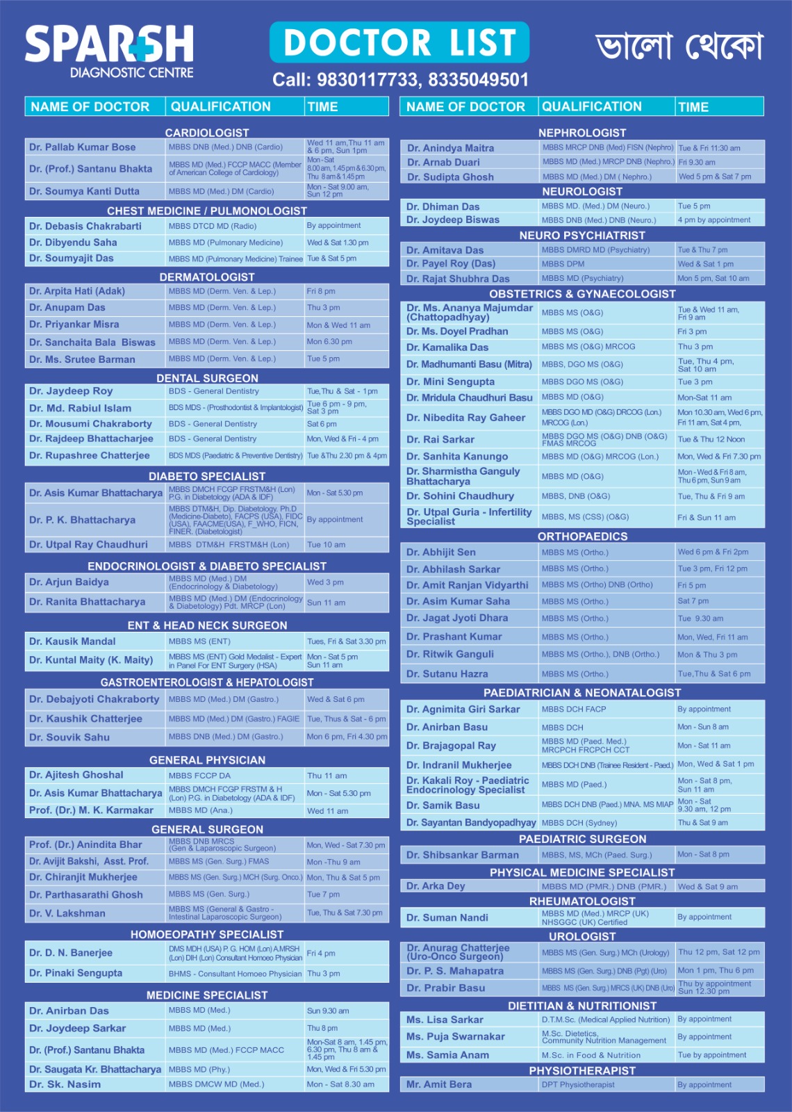
Sparsh Diagnostic Centre Doctor List
![]()


[…] 1. Obstructive Shock […]