Good circulation is essential for healthy legs and feet. Arteries carry oxygen-rich blood from the heart to the muscles and tissues, allowing them to function normally and heal after injury. When these arteries become narrowed or blocked, blood flow can decrease — sometimes gradually and without obvious symptoms.
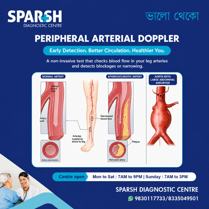
A peripheral arterial Doppler is a non-invasive ultrasound test that helps evaluate blood flow through the arteries of the arms or legs. It can identify areas where blood flow is reduced, arteries are narrowed, or an artery may be blocked. Doppler ultrasound is particularly useful when peripheral artery disease (PAD) is suspected.
PAD is usually associated with atherosclerosis, in which fatty deposits build up inside the arteries. It can cause leg pain while walking, cold feet, weak pulses and, in advanced cases, wounds that do not heal. Importantly, PAD is not only a problem affecting the legs. It is also a marker of wider cardiovascular disease and is associated with a higher risk of heart attack and stroke.
What is a peripheral arterial Doppler?
A peripheral arterial Doppler is an ultrasound examination used to assess blood flow through peripheral arteries, most commonly the arteries supplying the legs.
Unlike an X-ray or CT scan, it does not use radiation. A sonographer or radiologist uses an ultrasound probe over the skin to produce images of the blood vessels and assess the movement of blood within them.
The Doppler component provides information about the direction, speed and pattern of blood flow. Colour Doppler can help visualize blood movement, while spectral Doppler produces waveforms that allow the examiner to assess characteristics such as velocity, resistance and phasicity.
The examination can help detect:
- Narrowing or stenosis of an artery
- Partial arterial blockage
- Complete arterial occlusion
- Reduced blood flow
- Changes in Doppler waveforms caused by disease
- Abnormal blood flow beyond a narrowed segment
- Some complications following vascular procedures
Research has shown that Doppler ultrasound can be useful in assessing the severity and location of lower-limb peripheral arterial disease.
What is a peripheral Doppler test used for?
A peripheral Doppler test is mainly used to determine whether blood is flowing normally through the arteries supplying an arm or leg.
It may be recommended if a person has symptoms such as leg pain while walking, unexplained coldness of a foot, weak pulses, numbness, skin colour changes or wounds that heal slowly.
The test can also be useful for people with risk factors for vascular disease, including diabetes, high cholesterol, high blood pressure, smoking history and established cardiovascular disease.
One of the most common conditions investigated is peripheral artery disease, where narrowed arteries reduce blood flow to the limbs. Doppler ultrasound can help locate the affected artery and assess the characteristics of blood flow around the abnormal area.
However, peripheral arterial Doppler is not always the only test required. Doctors may also use an ankle-brachial index (ABI), toe-brachial index (TBI), segmental pressures or other vascular imaging depending on the symptoms and initial findings. Current guidelines recommend resting ABI as an important initial physiological test for suspected PAD.
How is a peripheral arterial Doppler performed?
The examination is relatively straightforward.
You will generally lie comfortably on an examination bed while the sonographer exposes the area being examined. A small amount of ultrasound gel is applied to the skin, and a handheld transducer is moved along the course of the arteries.
The sonographer may examine several arteries, depending on the reason for the test. In a lower-limb study, this can include arteries in the thigh, knee and lower leg.
The ultrasound system records both the appearance of the artery and the movement of blood through it. Spectral Doppler produces characteristic waveforms that can provide information about arterial resistance and blood-flow patterns.
The examination is painless and does not involve needles or injections. There is usually little or no preparation required, although specific instructions can vary depending on the centre and the type of vascular examination being performed.
What happens during a peripheral arterial Doppler?
The sonographer does more than simply look for a visible blockage.
Several features may be assessed, including:
Arterial wall and lumen: The ultrasound image can show the appearance of the vessel and, in some cases, plaque or other abnormalities.
Blood-flow velocity: Blood tends to accelerate as it passes through a narrowed section of an artery. Measuring velocity can therefore help identify significant stenosis.
Doppler waveform: The shape of the waveform provides important information about arterial function.
Flow beyond a blockage: Blood flow may become dampened or delayed downstream from a significant stenosis or occlusion.
Modern vascular Doppler reporting emphasizes descriptors such as flow direction, phasicity and resistance rather than relying on the older practice of simply calling every waveform “triphasic,” “biphasic” or “monophasic.”
What are normal arterial Doppler results?
A normal peripheral arterial Doppler generally shows good blood flow without significant narrowing or obstruction.
At rest, normal peripheral arterial waveforms are typically multiphasic, with rapid systolic acceleration, a sharp systolic peak and a component of reverse flow during diastole. A clear spectral window may also be present.
In simple terms, a reassuring report may describe:
- Patent arteries with no significant stenosis
- Normal or appropriate blood-flow velocities
- Multiphasic arterial waveforms
- No significant plaque-related narrowing
- No evidence of arterial occlusion
- Preserved distal blood flow
It is important to understand that there is no single “normal number” that applies to every artery and every patient. Doppler results are interpreted in relation to the particular vessel, the patient’s clinical situation, waveform characteristics and validated laboratory criteria.
For example, a biphasic or even monophasic waveform does not automatically mean severe PAD in every individual. Age, cardiac output, exercise, temperature and other physiological factors can influence waveform appearance.
What are abnormal Doppler results?
Abnormal Doppler findings can indicate that blood is not moving normally through an artery.
Depending on the location and severity of disease, a report may describe:
- Increased peak systolic velocity
- Significant spectral broadening
- Loss of the normal reverse-flow component
- Monophasic or dampened waveforms
- Delayed or prolonged systolic upstroke
- Reduced blood-flow velocity beyond a stenosis
- Very high velocities at a narrowed segment
- No detectable flow in an occluded artery
The Society for Vascular Medicine and Society for Vascular Ultrasound consensus statement notes that, in peripheral arterial disease, a velocity ratio greater than 2 can be associated with approximately 50–74% diameter stenosis, while a ratio greater than 4 can be associated with more severe stenosis. These thresholds must be interpreted within the appropriate vascular examination protocol and clinical context.
A significant narrowing can cause blood flow to accelerate through the narrowed area. Beyond the stenosis, the waveform may become dampened because the pressure and flow reaching the downstream artery have been affected.
A report showing an abnormal waveform does not, by itself, tell the entire story. The radiologist or vascular specialist considers the complete Doppler examination alongside symptoms, physical findings and other tests.
What are the three signs of peripheral artery disease?
There are several possible signs of PAD, but three particularly important symptoms are:
Leg pain while walking
This is one of the classic symptoms. Pain, aching or cramping — often in the calf — may occur after walking or climbing stairs and improve after resting. This is known as intermittent claudication.
Coldness or colour changes
One foot or lower leg may feel noticeably colder than the other. The skin may also appear pale, bluish or otherwise different in colour when circulation is significantly reduced.
Slow-healing wounds
Poor arterial circulation can make it difficult for tissues to receive sufficient oxygen and nutrients. As a result, cuts or sores on the toes, feet or legs may take unusually long to heal.
Other possible signs include weak pulses, numbness, leg weakness, shiny skin, reduced hair growth and changes in toenail growth.
Some people with PAD have few or no obvious symptoms, which is one reason risk-factor assessment and appropriate vascular testing are important.
What are the 5 P’s of peripheral artery disease?
The commonly taught 5 P’s are:
- Pain – severe or sudden limb pain
- Pallor – unusual paleness of the affected limb
- Pulselessness – a weak or absent pulse
- Paresthesia – numbness or abnormal sensation
- Paralysis – weakness or inability to move the limb
These signs are particularly associated with acute limb ischemia, which is a sudden reduction or interruption of arterial blood flow and should be treated as a medical emergency.
The 5 P’s should not be confused with the more common symptoms of chronic PAD. A person does not need to have all five signs to have peripheral artery disease.
Sudden severe leg pain accompanied by a cold, pale foot, numbness, weakness or loss of pulse requires urgent medical attention.
What causes peripheral artery disease?
The most common cause is atherosclerosis, where cholesterol and other substances accumulate within artery walls and form plaques.
Risk factors include:
- Smoking or tobacco use
- Diabetes
- High cholesterol
- High blood pressure
- Increasing age
- Obesity
- Physical inactivity
- A history of cardiovascular disease
PAD and coronary artery disease share many of the same risk factors. Having PAD therefore warrants attention not only to circulation in the legs but also to overall cardiovascular health.
What is the best treatment for peripheral artery disease?
There is no single treatment that is best for every person with PAD. Treatment depends on whether the disease is mild, symptomatic, severe or threatening the limb.
For many patients, treatment begins with risk-factor management, structured exercise and appropriate medication.
Doctors may recommend:
- Stopping smoking and tobacco use
- Structured exercise therapy
- Cholesterol-lowering treatment, particularly statin therapy
- Blood-pressure management
- Diabetes management
- Antiplatelet or other antithrombotic therapy when appropriate
- Weight and dietary management
- Regular foot care
The 2024 ACC/AHA guideline emphasizes structured exercise as a core component of PAD care and recommends evidence-based medical therapy to reduce cardiovascular and limb-related complications.
For patients whose symptoms remain significantly limiting despite medical treatment and structured exercise, revascularization may be considered. Depending on the anatomy and severity of disease, this may involve angioplasty, stenting, bypass surgery or another vascular procedure.
In chronic limb-threatening ischemia, restoring blood flow may be necessary to prevent tissue loss and limb amputation.
What is the best natural treatment for peripheral artery disease?
There is no natural remedy that can reliably remove an established arterial blockage. Be cautious about products or supplements claiming to “clean” or “open” arteries without medical treatment.
However, several lifestyle measures can naturally support cardiovascular and vascular health.
Regular physical activity is one of the most important. Structured walking or exercise programs can improve walking ability and reduce PAD symptoms. Exercise should be planned according to the severity of disease and individual medical circumstances.
Stopping smoking is equally important. Tobacco damages blood vessels and can worsen PAD and its complications.
Eating a heart-healthy diet is another valuable step. Focus on vegetables, fruits, whole grains, legumes, nuts and other minimally processed foods while limiting excessive saturated fat, salt and added sugar.
Maintaining a healthy weight and controlling blood pressure, cholesterol and blood glucose can also reduce cardiovascular risk.
These lifestyle measures are best viewed as part of medical treatment rather than replacements for it.
When should you consider a peripheral arterial Doppler?
You may be advised to undergo a peripheral arterial Doppler if you experience:
- Repeated calf, thigh or buttock pain while walking
- Leg pain that improves with rest
- A cold foot or lower leg
- Weak or difficult-to-feel pulses
- Numbness or weakness in a limb
- Changes in skin colour
- Slow-healing wounds on the foot or leg
- Unexplained changes in the appearance of the legs or feet
People with significant risk factors such as diabetes, smoking history or known cardiovascular disease may also need vascular assessment based on their individual risk and symptoms.
Why early detection matters
Peripheral artery disease can be easy to overlook because symptoms may initially be mild. Some people simply assume that leg pain or reduced walking ability is part of getting older.
But reduced circulation should not automatically be attributed to age.
Early diagnosis gives patients and doctors an opportunity to address risk factors, improve walking ability and reduce the chance of serious complications. It also provides an opportunity to look at the person’s broader cardiovascular health.
A peripheral arterial Doppler is therefore more than an imaging test. It can provide valuable information about how effectively blood is reaching the tissues of the limbs.
Frequently asked questions
Is peripheral arterial Doppler painful?
No. It is a non-invasive ultrasound examination. A transducer is moved over the skin with ultrasound gel. There are no needles or injections involved in a routine Doppler study.
Can a Doppler detect a blocked artery?
Yes. Doppler ultrasound can identify abnormal or absent blood flow and can help locate areas of narrowing or occlusion. It can also assess the blood-flow changes associated with arterial stenosis.
Is peripheral arterial Doppler the same as an ABI test?
No. They are different tests, although they may be used together. ABI compares blood pressure at the ankle with blood pressure in the arm and is an important physiological test for PAD. Doppler ultrasound directly evaluates arterial anatomy and blood-flow characteristics.
Can a normal Doppler rule out all circulation problems?
Not necessarily. Test performance depends on the vessels examined, the disease being investigated and technical factors. Some patients may need ABI, TBI, exercise testing or additional imaging if symptoms persist despite an initially reassuring examination.
Does a monophasic waveform always mean severe PAD?
No. A monophasic waveform can be associated with arterial disease, particularly when it is accompanied by other abnormal findings, but waveform interpretation must consider the location, clinical context and other Doppler parameters. Age, cardiac function and other physiological factors can also affect waveforms.
Can peripheral artery disease be cured naturally?
Lifestyle changes can significantly improve cardiovascular health and PAD symptoms, but there is no proven natural treatment that can reliably dissolve an established arterial blockage. Smoking cessation, structured exercise and a heart-healthy diet are important components of evidence-based PAD care.
What should I do if my foot suddenly becomes cold, pale and painful?
Seek urgent medical attention. Sudden severe pain, pallor, loss of pulse, numbness or weakness can indicate acute limb ischemia, which requires immediate assessment and treatment.
Final thoughts
A peripheral arterial Doppler is a safe and useful way to evaluate blood flow through the arteries of the limbs. It can help identify narrowing, blockages and abnormal blood-flow patterns and can play an important role in the assessment of peripheral artery disease.
If you have persistent leg pain while walking, cold feet, changes in skin colour, weak pulses or wounds that are slow to heal, don’t simply ignore the symptoms. Early evaluation can help identify circulation problems and guide appropriate treatment.
At Sparsh Diagnostic Centre, a peripheral arterial Doppler can help assess arterial blood flow and identify possible blockages or narrowing. The centre is open Monday to Saturday from 7 AM to 9 PM and Sunday from 7 AM to 3 PM. For appointments and enquiries, you can contact 9830117733 / 8335049501.
This article is intended for general health education and does not replace an examination or advice from a qualified doctor. Doppler findings should always be interpreted by an appropriately qualified medical professional in the context of the patient’s symptoms and other investigations.
Book Your Peripheral Arterial Doppler Test Today!
📍 Visit Sparsh Diagnostic Centre
🕒 Mon–Sat: 7 AM to 9 PM | Sun: 7 AM to 3 PM
📞 Call/WhatsApp: 9830117733 / 8335049501
🌐 www.sparshdiagnostica.com
#BhaloTheko
Disclaimer:
No content on this site, regardless of date, should ever be used as a substitute for direct medical advice from your doctor or other qualified clinician.