
Medical imaging has revolutionized how doctors diagnose and treat diseases. Among the most advanced and widely used imaging techniques is Magnetic Resonance Imaging (MRI). Unlike X-rays or CT scans, MRI uses powerful magnets and radio waves to produce detailed images of organs, tissues, and bones — all without exposing the patient to harmful ionizing radiation.
Whether it’s used for brain scans, spine problems, sports injuries, or detecting tumors, MRI has become a gold standard in diagnostic imaging. In this blog, we’ll explore what MRI is, how it works, the types of MRI scans available, its benefits, risks, and what patients can expect before, during, and after the procedure.
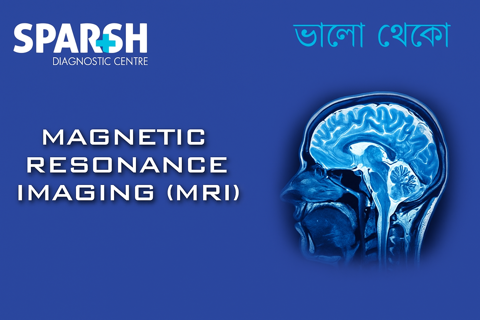
What is an MRI?
Magnetic Resonance Imaging (MRI) is a non-invasive imaging test that uses magnetic fields and radio waves to create highly detailed images of internal structures. It is particularly useful for evaluating soft tissues, such as the brain, spinal cord, muscles, ligaments, and blood vessels, which may not be as visible on X-rays or CT scans.
MRI does not use radiation, making it a safer choice for patients who require multiple scans over time.
How Does MRI Work?
MRI technology is based on the principles of nuclear magnetic resonance (NMR):
Magnetism: The MRI machine contains a large magnet that aligns the hydrogen protons in the body.
Radio Waves: Short bursts of radiofrequency waves are sent into the body, temporarily disrupting the alignment of these protons.
Signal Detection: As the protons return to their original position, they release energy.
Image Formation: The scanner detects this energy and a computer processes it into detailed, cross-sectional images.
Different tissues in the body (e.g., fat, muscle, brain matter) respond differently, allowing radiologists to distinguish between healthy and diseased tissue.
Types of MRI Scans
There are several specialized MRI scans, each tailored to specific parts of the body or medical conditions:
Brain MRI – Detects tumors, strokes, multiple sclerosis, aneurysms, and infections.
Spinal MRI – Used to assess herniated discs, spinal stenosis, and nerve compression.
Musculoskeletal MRI – Evaluates sports injuries, ligament tears, and joint problems.
Cardiac MRI – Provides detailed images of the heart’s structure and blood flow.
Breast MRI – Complements mammography for high-risk patients or dense breast tissue.
Functional MRI (fMRI) – Measures brain activity by detecting blood flow changes.
Magnetic Resonance Angiography (MRA) – Focuses on blood vessels to detect aneurysms or blockages.
Why is MRI Done?
MRI scans are performed to help doctors diagnose, monitor, or rule out various health conditions. Common reasons include:
Musculoskeletal injuries (torn ligaments, cartilage damage).
Cancer detection and staging.
Cardiovascular diseases (heart disease, blocked arteries).
Spinal cord problems (disc herniation, nerve compression).
Infections or inflammatory diseases.
Benefits of MRI
MRI offers several advantages over other imaging techniques:
No radiation exposure – safer than CT scans or X-rays.
Detailed soft tissue imaging – especially useful for the brain, muscles, and organs.
Non-invasive – no surgery required.
Versatile applications – from head to toe imaging.
Early detection of diseases – allows for timely treatment.
Risks and Limitations of MRI
Although MRI is generally safe, there are certain risks and limitations:
Claustrophobia – Some patients feel anxious inside the MRI machine.
Noise – The machine produces loud knocking sounds, though earplugs or headphones are provided.
Magnet-related risks – People with pacemakers, cochlear implants, or certain metal implants may not be eligible.
Contrast dye risks – Rare allergic reactions or kidney complications may occur with gadolinium contrast.
Time-consuming – Scans usually last 30–60 minutes, longer than CT scans.
How to Prepare for an MRI Scan
Preparation may vary depending on the type of MRI, but common steps include:
Inform your doctor if you have metal implants, pacemakers, or pregnancy.
Remove all metallic objects, including jewelry, watches, hearing aids, and belts.
Wear comfortable clothing or change into a hospital gown.
Fasting may be required for certain abdominal MRIs.
Contrast dye may be injected depending on the scan type.
What Happens During an MRI?
The patient lies on a motorized table that slides into the MRI machine.
Straps and cushions may be used to minimize movement.
The technician operates the machine from another room, while staying in communication via microphone.
The scan produces loud tapping or thumping noises — headphones or earplugs are provided.
Patients must stay very still to ensure clear images.
The scan is painless, though some may feel discomfort due to stillness or anxiety in confined spaces.
After the MRI
Patients can typically resume normal activities immediately.
If contrast dye was used, drinking water helps flush it out.
Results are analyzed by a radiologist and shared with your doctor.
MRI vs. CT Scan: What’s the Difference?
| Feature | MRI | CT Scan |
|---|---|---|
| Technology | Magnetic fields & radio waves | X-rays |
| Radiation Exposure | None | Yes |
| Best for | Soft tissues (brain, muscles, organs) | Bones, chest, trauma cases |
| Duration | 30–60 minutes | 5–15 minutes |
| Cost | Generally higher | Lower |
Cost of MRI in India
The cost of an MRI scan in India can range between ₹3,000 to ₹25,000, depending on:
Type of MRI (brain, spine, cardiac, etc.)
Use of contrast dye
Diagnostic center or hospital facilities
City or region
FAQs About MRI
1. Is an MRI painful?
No, an MRI is a painless procedure. Some patients may experience mild discomfort from lying still or claustrophobia.
2. How long does an MRI scan take?
Most MRI scans take between 30 to 60 minutes, depending on the body part being examined.
3. Can everyone have an MRI?
No, people with pacemakers, certain implants, or metallic fragments in the body may not be eligible.
4. Do I need to fast before an MRI?
Not always. Fasting is usually required only for abdominal or pelvic MRIs.
5. Is MRI safe during pregnancy?
MRI is generally safe in pregnancy, especially after the first trimester. However, contrast dye is usually avoided unless absolutely necessary.
6. What is the difference between MRI with and without contrast?
Contrast-enhanced MRI provides more detailed images, especially for detecting tumors, blood vessels, and infections.
7. How soon will I get my results?
Results are typically available within 24 to 48 hours, depending on the diagnostic centre.
Magnetic Resonance Imaging (MRI) is one of the most powerful tools in modern medicine, offering detailed, radiation-free images that help diagnose a wide range of conditions. While the procedure may feel intimidating to some due to its noise and enclosed space, it is a safe, painless, and highly effective diagnostic test.
If your doctor has recommended an MRI, rest assured that it is a step toward accurate diagnosis and effective treatment planning.
#BhaloTheko
Disclaimer:
No content on this site, regardless of date, should ever be used as a substitute for direct medical advice from your doctor or other qualified clinician.

![]()


[…] MRI Brain and Brainstem – to identify strokes, tumors, demyelination […]
[…] MRI […]
[…] CT or MRI (if […]
[…] scan or MRI – detailed liver […]
[…] or MRI to assess liver, spleen, and brain […]
[…] Scan or MRI: Detects cirrhosis, varices, thrombosis, and […]
[…] MRI (Magnetic Resonance Imaging): Confirms the diagnosis and reveals associated injuries to menisci, cartilage, or other ligaments. […]
[…] MRI or CT […]
[…] 4. MRI of Spine or Pelvis […]
[…] MRI/MR angiography: Offers high-resolution images of abdominal vessels. […]
[…] MRI or CT scans for arterial clots […]
[…] scans or MRIs may be used if internal bleeding or organ damage is […]
[…] 3. MRI […]
[…] MRI or CT scans may be used to rule out herniated discs or tumors. […]
[…] and even those leading sedentary lifestyles. While traditional imaging methods like X-rays and MRIs are widely used, musculoskeletal ultrasound (MSK ultrasound) has emerged as a powerful, […]
[…] 6. MRI Abdomen […]
[…] Scan/MRI: More detailed […]
[…] MRI: Detects soft tissue damage and inflammation. […]
[…] or MRI Brain – to rule out structural brain damage or […]
[…] CT/MRI: Used when malignancy or structural abnormalities are suspected. […]
[…] MRI (to check tissue swelling) […]
[…] 3. Imaging (X-Ray or MRI) […]
[…] CT or MRI (in rare […]
[…] CT/MRI: If obstructive causes or complications are suspected […]
[…] MRI/MRCP: Offers detailed ductal anatomy. […]
[…] X-rays, MRI, or ultrasound to evaluate joint […]
[…] MRI or CT Scans: Imaging tests to detect structural issues in the muscles or nervous system. […]
[…] 6. Imaging (MRI/Ultrasound) […]
[…] Diagnostic imaging (X-rays, USG, CT, MRI) […]
[…] 4. Ultrasound or MRI […]
[…] 1. Magnetic Resonance Imaging (MRI) […]
[…] MRI or CT Scans: Rule out other conditions with similar symptoms. […]
[…] Tests: MRI or CT scans may be ordered to rule out other causes of sciatic nerve […]
[…] X-rays and MRI scans can reveal cartilage loss and joint […]
[…] MRI: Detects cartilage damage and soft tissue inflammation. […]
[…] X-rays, MRI, and ultrasound to detect joint erosion and […]
[…] or MRI for fat […]
[…] MRI (Magnetic Resonance Imaging): Provides detailed images of soft tissues, discs, and nerves. […]
[…] Tests (MRI/CT): To check for neurological or structural […]
[…] Scan or MRIUsed to detect TB in organs other than the […]
[…] or MRI: Helps detect inflammation in joints or soft […]
[…] 3. Magnetic Resonance Imaging (MRI) of the Brain […]
[…] MRI (Magnetic Resonance Imaging): Shows soft tissue details including discs, nerves, and spinal cord. […]
[…] Ultrasound/MRI: Imaging to detect structural abnormalities like masses or swelling. […]
[…] 2. MRI (Magnetic Resonance Imaging) […]
[…] CT scan […]
[…] 2. MRI Scan […]
[…] MRI scan – for a detailed view of the uterus and ovaries. […]
[…] imaging like CT scans, MRIs, and ultrasounds can detect internal issues […]
[…] tests, urine analysis, and imaging (like CT or MRI) may be needed to identify underlying causes such as infection, metabolic imbalance, or brain […]
[…] MRI Venography […]
[…] MRI or CT […]