


If you have persistent joint pain, a sports injury, a suspected tendon tear, muscle strain, swelling, or a problem that seems difficult to pinpoint, your doctor may recommend a musculoskeletal ultrasound, often called an MSK ultrasound.
Unlike a standard ultrasound that may be used to look at organs such as the abdomen, pelvis or thyroid, an MSK ultrasound is specifically designed to examine the muscles, tendons, ligaments, nerves, joints and other soft tissues involved in movement.
One of its biggest advantages is that it does not just provide a still picture. The person performing the examination can watch structures in real time and while they move. This can be particularly helpful when pain or an abnormality only becomes apparent during movement.
MSK ultrasound is also non-invasive, does not use radiation and generally requires little or no preparation. A typical examination takes around 15 to 30 minutes, although the exact time depends on the area being examined and whether additional assessment or an ultrasound-guided procedure is required.
What is a musculoskeletal ultrasound?
A musculoskeletal ultrasound is a diagnostic imaging test that uses high-frequency sound waves to produce real-time images of the body’s musculoskeletal structures.
During the examination, a small handheld device called a transducer or probe is placed against the skin. A layer of ultrasound gel helps the sound waves travel efficiently between the probe and the body.
The sound waves bounce differently from different tissues. The ultrasound machine processes these returning signals and creates images that the radiologist or trained healthcare professional can interpret.
An MSK ultrasound can examine structures such as:
- Muscles
- Tendons
- Ligaments
- Nerves
- Joints and joint spaces
- Bursae
- Superficial soft tissues
- Fluid collections
- Some cysts and soft-tissue masses
It can be used to investigate problems affecting the shoulder, elbow, wrist, hand, hip, knee, ankle and foot, as well as other areas of the body.
For example, an MSK ultrasound may be recommended when a doctor suspects tendinitis, a tendon tear, muscle injury, bursitis, ligament injury, nerve entrapment or joint inflammation.
What is the difference between an ultrasound and an MSK ultrasound?
This is a common source of confusion because an MSK ultrasound is still an ultrasound examination.
The main difference is what is being examined and how the examination is performed.
A general ultrasound is a broad imaging technique used for many parts of the body. Depending on the examination, it may be used to assess organs such as the liver, gallbladder, kidneys, uterus, ovaries or thyroid.
An MSK ultrasound is a specialised ultrasound examination focused on the musculoskeletal system. The person performing the scan uses appropriate probes and scanning techniques to assess muscles, tendons, ligaments, nerves, joints and related soft tissues.
Another important difference is the emphasis on dynamic assessment. During an MSK examination, you may be asked to move your arm, leg, joint or another body part while the ultrasound images are being observed. This can help reveal abnormalities that are difficult to appreciate when the body part is completely still.
So, when people ask, “What is the difference between ultrasound and musculoskeletal ultrasound?” the simplest answer is:
MSK ultrasound is a specialised form of diagnostic ultrasound designed specifically to evaluate the muscles, tendons, ligaments, nerves, joints and other structures involved in movement.
What will a musculoskeletal ultrasound show?
One of the strengths of MSK ultrasound is its ability to provide detailed images of many superficial soft-tissue structures.
Depending on the area being examined, it may help identify:
- Tendon inflammation or tendinitis
- Partial or complete tendon tears
- Muscle strains and tears
- Ligament sprains or tears
- Bursitis
- Joint fluid or effusions
- Inflammatory changes
- Nerve entrapment
- Ganglion cysts
- Certain soft-tissue masses
- Foreign bodies in superficial soft tissue
- Changes associated with some inflammatory joint conditions
It can also be useful for examining blood flow with Doppler ultrasound when vascular or inflammatory activity needs to be assessed.
For example, if you have shoulder pain, an MSK ultrasound can assess structures such as the rotator cuff tendons and surrounding soft tissues. For heel or ankle pain, it may help evaluate the Achilles tendon. In the wrist, it can be useful when conditions such as carpal tunnel syndrome are suspected.
The exact usefulness of the test depends on the symptoms, suspected diagnosis and area being examined.
How do I prepare for an MSK ultrasound?
One of the convenient things about musculoskeletal ultrasound is that there is usually little to no special preparation.
In most cases, you can eat, drink and take your usual medications as normal unless your doctor or diagnostic centre has given you different instructions.
Before your appointment:
- Wear loose, comfortable clothing.
- Make sure the area being examined can be easily exposed.
- Avoid wearing jewellery around the area if possible.
- Bring previous imaging reports or relevant medical records if your doctor has asked for them.
- Follow any specific instructions provided by the diagnostic centre.
You generally do not need fasting for a routine MSK ultrasound. However, if your examination is being combined with another test or a specific procedure, you may receive additional instructions.
During the scan, a small amount of gel is placed on the skin and the probe is moved over the area being examined. You may also be asked to move the affected joint or limb.
How long does a musculoskeletal ultrasound take?
Most musculoskeletal ultrasound examinations take approximately 15 to 30 minutes. Some examinations may take longer depending on the area being assessed, the complexity of the problem, or whether multiple structures need to be examined.
For example, examining one relatively small area may be quicker than assessing several tendons, muscles or joints.
If an ultrasound-guided injection, aspiration or another intervention is performed during the same appointment, additional time will be needed.
The good news is that you normally do not need recovery time after a routine diagnostic MSK ultrasound. You can generally return to your usual activities immediately afterwards.
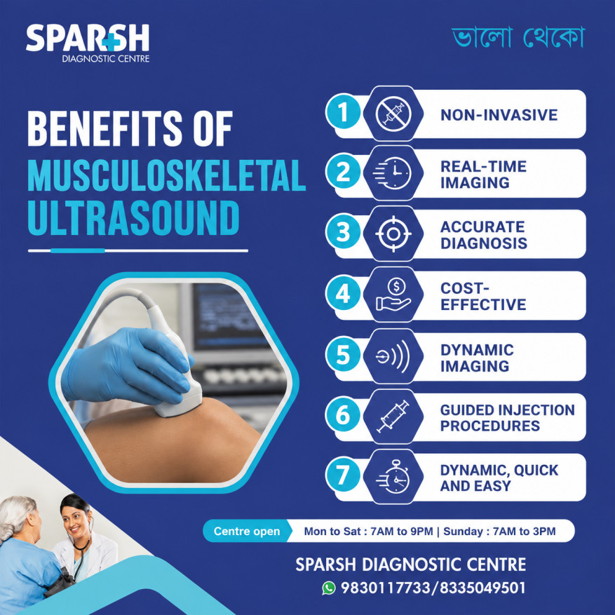
What are the benefits of musculoskeletal ultrasound?
There are several reasons why MSK ultrasound has become an important tool in orthopaedics, sports medicine, rheumatology, rehabilitation and pain management.
1. It is non-invasive
A standard diagnostic MSK ultrasound does not require needles or injections. The probe simply moves over the skin.
2. It does not use radiation
Ultrasound uses sound waves rather than X-rays. This makes it particularly useful when repeated imaging is needed.
3. It provides real-time imaging
This is perhaps its most distinctive advantage.
The examiner can watch tendons, muscles and joints as they move. If a patient experiences pain during a particular movement, the area can sometimes be assessed while that movement is being performed.
4. It is relatively quick
Most examinations take around 15 to 30 minutes.
5. It can be more accessible and cost-effective
Ultrasound equipment is generally more accessible than MRI, and MSK ultrasound is usually less expensive than MRI. This can make it useful when appropriate for the clinical question.
6. It can assess superficial structures in considerable detail
High-frequency ultrasound probes can provide excellent images of superficial tendons, nerves, muscles and other soft tissues. Modern systems can provide very high-resolution imaging of small structures.
7. It can guide procedures
Ultrasound allows the healthcare professional to see the target and needle in real time during certain procedures. This can be useful for injections, aspirations and other interventions.
What’s the difference between MRI and MSK ultrasound?
MRI and MSK ultrasound are both valuable imaging tests, but they provide different types of information.
MSK ultrasound is particularly useful for superficial soft tissues and dynamic assessment. The examiner can watch a tendon or muscle while it moves, compare one side with the other and focus directly on the area where the patient feels pain.
MRI, on the other hand, provides detailed cross-sectional images and is particularly valuable for deeper structures, bone marrow and structures within joints. Yale Medicine notes that MRI is generally preferred when evaluating structures deep within a joint.
A simple comparison looks like this:
| Feature | MSK ultrasound | MRI |
|---|---|---|
| Radiation | No | No |
| Real-time movement | Yes | Generally no |
| Superficial tendons | Excellent | Excellent |
| Dynamic assessment | Excellent | Limited |
| Deep joint structures | More limited | Excellent |
| Bone marrow | Limited | Excellent |
| Examination time | Usually 15–30 minutes | Usually longer |
| Claustrophobia | Not an issue | Can be an issue |
| Image-guided injections | Can guide in real time | Less commonly used for routine guidance |
Neither test is automatically “better.” The right choice depends on what your doctor needs to see.
Which is better for hip pain, ultrasound or MRI?
There is no single answer because hip pain can come from several different structures.
An MSK ultrasound can be particularly useful for assessing superficial tendons, bursae, muscles and other soft tissues around the hip. It can also provide dynamic information and can be used to guide certain injections when appropriate.
MRI is generally more useful when the concern involves deep structures within the hip joint, bone marrow or other areas that ultrasound cannot adequately visualise. Ultrasound has inherent limitations because sound waves do not penetrate bone well and deeper structures can be difficult to assess.
So, for someone with hip pain, the better test depends on the suspected cause.
Your doctor may recommend ultrasound first when a superficial tendon or bursal problem is suspected, while MRI may be preferred when a deeper joint or bone-related problem needs investigation.
What is the role of musculoskeletal ultrasound in pain medicine?
MSK ultrasound has become increasingly useful in pain medicine because it can help doctors identify the structure responsible for pain and guide certain treatments with real-time imaging.
For example, ultrasound can help identify inflammation around a tendon or bursa, joint fluid, nerve abnormalities or other soft-tissue problems.
It can also guide procedures such as selected joint, tendon, bursal or nerve injections. Instead of relying only on anatomical landmarks, the clinician can see the target and needle during the procedure.
Research has generally found that ultrasound-guided injections are more accurate than landmark-guided injections, although the degree of improvement in pain relief and function varies according to the procedure and condition. A 2024 umbrella review found particularly useful evidence for ultrasound guidance in areas including the hip and knee.
This makes MSK ultrasound useful not only for diagnosis but also as part of an overall pain assessment and treatment strategy.
What happens during a musculoskeletal ultrasound?
The process is usually straightforward.
You will be positioned either sitting or lying down, depending on the part of the body being examined.
The sonographer or radiologist applies ultrasound gel to the skin. The handheld probe is then gently moved over the area of concern.
You may be asked questions about where you feel pain and may be asked to move the affected body part.
For example, if you have shoulder pain, you may be asked to raise or rotate your arm. If the examination involves the knee or ankle, you may be asked to move the joint in particular directions.
This dynamic component is one of the reasons MSK ultrasound can be so useful. The examiner is not limited to looking at the tissues while they are completely still.
The scan itself is generally painless, although you may feel some pressure if the probe is placed over a particularly tender area.
Are there any limitations to musculoskeletal ultrasound?
Although MSK ultrasound has many advantages, it is not a replacement for every other imaging test.
Ultrasound has difficulty penetrating bone, so it cannot provide the same information about the internal structure of bones as MRI. Deep structures may also be difficult to visualise, particularly when they are located beneath substantial amounts of tissue.
The quality of the examination can also depend on the equipment and the experience of the person performing and interpreting the scan.
For this reason, your doctor may recommend an X-ray, MRI, CT scan or another test if ultrasound does not answer the clinical question.
In many cases, ultrasound and MRI are not competing tests. They are complementary tools that can be used at different stages of diagnosis.
Why might your doctor recommend an MSK ultrasound?
An MSK ultrasound may be recommended when you have:
- Persistent muscle or joint pain
- A suspected tendon injury
- A sports-related injury
- Swelling around a joint
- Suspected bursitis
- A muscle strain or tear
- Possible ligament injury
- Numbness or symptoms suggesting nerve entrapment
- A soft-tissue lump or swelling
- An inflammatory musculoskeletal condition
- A need for an ultrasound-guided procedure
It can also be used to monitor certain conditions over time and assess changes during treatment.
Frequently asked questions about musculoskeletal ultrasound
1. What is a musculoskeletal ultrasound?
A musculoskeletal ultrasound is a specialised ultrasound examination used to evaluate muscles, tendons, ligaments, nerves, joints and other soft tissues. It uses sound waves rather than radiation and can provide real-time images, including images while the body part is moving.
2. How do I prepare for an MSK ultrasound?
Usually, no special preparation is required. Wear loose, comfortable clothing that allows easy access to the area being examined. You generally do not need to fast for a routine MSK ultrasound. Follow any specific instructions provided by your doctor or diagnostic centre.
3. What’s the difference between MRI and MSK ultrasound?
MSK ultrasound provides real-time imaging and is particularly useful for superficial soft tissues and structures during movement. MRI provides detailed cross-sectional images and is generally better for deep joint structures, bone marrow and areas that ultrasound cannot adequately reach.
4. How long does a musculoskeletal ultrasound take?
A routine MSK ultrasound usually takes around 15 to 30 minutes, although a more complex examination or an ultrasound-guided procedure may take longer.
5. What is the difference between an ultrasound and an MSK ultrasound?
Ultrasound is the broader imaging technology. MSK ultrasound is a specialised application of that technology specifically designed to examine the musculoskeletal system, including muscles, tendons, ligaments, nerves and joints.
6. Which is better for hip pain, ultrasound or MRI?
It depends on the suspected cause of the hip pain. Ultrasound can be very useful for superficial tendons, bursae and other soft tissues and can provide dynamic assessment. MRI is generally more suitable for deeper structures inside the hip joint and bone-related abnormalities.
7. What are the benefits of musculoskeletal ultrasound?
Major benefits include no radiation, real-time imaging, dynamic assessment, relatively quick examination times, good visualisation of many superficial soft tissues and the ability to guide certain procedures. It is also generally accessible and cost-effective compared with MRI.
8. What is the role of musculoskeletal ultrasound in pain medicine?
It can help identify abnormalities associated with pain and can guide certain injections and other procedures. Real-time visualisation can improve the accuracy of needle placement and help the clinician avoid nearby structures such as nerves and blood vessels.
9. How do I prepare for a musculoskeletal ultrasound?
For most MSK ultrasound examinations, you simply need to wear comfortable clothing and make sure the area can be accessed easily. There is generally no fasting or other special preparation unless you have been given specific instructions.
10. What will a musculoskeletal ultrasound show?
It can show many abnormalities involving muscles, tendons, ligaments, nerves, bursae and joints, including tendon tears, tendinitis, muscle injuries, bursitis, fluid collections, nerve entrapment and some soft-tissue masses.
11. What is the difference between ultrasound and musculoskeletal ultrasound?
A standard ultrasound can be used to examine many different parts of the body. Musculoskeletal ultrasound is specifically performed to assess structures involved in movement, such as muscles, tendons, ligaments, nerves and joints. It often includes dynamic assessment while the patient moves.
Book a musculoskeletal ultrasound in Kolkata
If you have ongoing muscle, tendon, ligament or joint pain, getting the right imaging test can be an important step toward finding the cause.
A musculoskeletal ultrasound offers a convenient, radiation-free way to examine many soft-tissue problems in real time. It can be particularly useful when the suspected abnormality involves a tendon, muscle, ligament, nerve, bursa or superficial joint structure.
At Sparsh Diagnostic Centre, musculoskeletal ultrasound is available for the evaluation of a range of soft-tissue and musculoskeletal concerns.
Centre timings: Monday to Saturday, 7:00 AM to 9:00 PM; Sunday, 7:00 AM to 3:00 PM.
For appointments and enquiries: 9830117733 / 8335049501.
If your doctor has recommended an MSK ultrasound, bring your prescription and any previous imaging reports with you. The radiologist can then interpret the findings in the context of your symptoms and clinical history.
Important: An ultrasound report should always be interpreted alongside your symptoms and physical examination. If ultrasound cannot adequately answer the clinical question, your doctor may recommend additional imaging such as an MRI or X-ray.
Book your musculoskeletal ultrasound today!
📍 Visit Sparsh Diagnostic Centre
🕒 Mon–Sat: 7 AM to 9 PM | Sun: 7 AM to 3 PM
📞 Call/WhatsApp: 9830117733 / 8335049501
🌐 www.sparshdiagnostica.com
#BhaloTheko
Disclaimer:
No content on this site, regardless of date, should ever be used as a substitute for direct medical advice from your doctor or other qualified clinician.




[…] Musculoskeletal Ultrasound is particularly useful and non-invasive for detecting median nerve swelling. […]