


When a doctor recommends an ultrasound of the pelvis, you may be surprised to hear that there is more than one way to perform the examination. Two of the most commonly used approaches are transvaginal ultrasound (TVS) and transabdominal ultrasound.
Both use sound waves to create images of the reproductive organs, and both are safe, non-invasive imaging techniques. However, they are performed differently and can provide different levels of detail depending on what your doctor needs to see.
A transabdominal ultrasound uses a probe placed over the lower abdomen, while a transvaginal ultrasound uses a slender probe inserted gently into the vagina. In some situations, doctors may recommend both because the two approaches complement each other. The American Institute of Ultrasound in Medicine notes that more than one approach may be necessary for a complete evaluation of the female pelvis.
So, which is better: TVS or USG? And which ultrasound is best for examining the uterus?
The answer depends on what needs to be evaluated, the stage of pregnancy if you are pregnant, your symptoms, and your individual circumstances. There is no single ultrasound that is always better for everyone.
What is a transabdominal ultrasound?
A transabdominal ultrasound, sometimes simply called an abdominal or pelvic ultrasound, is performed by moving an ultrasound transducer over the skin of the lower abdomen.
A layer of ultrasound gel is applied to the skin to help the sound waves travel efficiently between the transducer and the body. The sonographer then moves the probe over the abdomen to obtain images of the uterus, ovaries, bladder and other pelvic structures.
For a pelvic transabdominal ultrasound, you are often asked to arrive with a comfortably full bladder. A full bladder can help push the bowel away and provide an acoustic window through which the pelvic organs can be viewed more clearly.
One of the biggest advantages of this approach is that it provides a broader view of the pelvis without inserting anything into the vagina.
What is a transvaginal ultrasound?
A transvaginal ultrasound, commonly known as TVS, uses a specially designed slender ultrasound probe that is gently inserted into the vagina.
The transducer is covered with a protective sheath and lubricated before insertion. The examination is usually performed with an empty bladder. The probe is positioned close to the uterus, cervix and ovaries, allowing the sonographer to obtain detailed images of these structures.
Because the transducer is much closer to the pelvic organs, TVS can often provide high-resolution images of structures that may be more difficult to assess in detail through the abdominal wall.
TVS is frequently used for evaluating the uterus and endometrium, ovarian abnormalities, pelvic pain, abnormal bleeding, infertility and certain early pregnancy concerns.
Transvaginal ultrasound vs abdominal ultrasound: What is the difference?
The main difference is where the ultrasound probe is placed.
| Feature | Transabdominal ultrasound | Transvaginal ultrasound |
|---|---|---|
| Probe location | Over the lower abdomen | Inside the vagina |
| Bladder | Usually full | Usually empty |
| Image detail | Good overall pelvic view | Often more detailed view of pelvic organs |
| Invasiveness | External examination | Internal examination |
| Uterus and endometrium | Can be evaluated | Often provides greater detail |
| Ovaries | Can be evaluated | Often provides detailed assessment |
| Early pregnancy | Useful depending on gestational age | Often particularly useful early in pregnancy |
| Patient experience | Generally comfortable | May cause mild pressure or discomfort |
| Field of view | Broader overview | Closer, detailed view |
The choice is not necessarily either-or. Johns Hopkins Medicine explains that pelvic ultrasound may be performed using either method or both, depending on the reason for the examination and the information required.
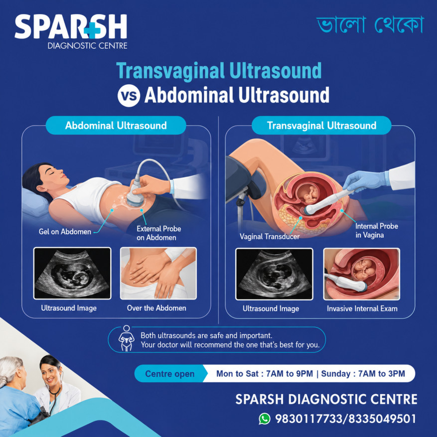
Which is better, TVS or USG?
This is one of the most common questions patients ask, but there isn’t a universal winner.
TVS is often better when detailed imaging of the uterus, endometrium, cervix or ovaries is required, because the transducer is positioned closer to these structures.
On the other hand, transabdominal ultrasound can be preferable when a broader view of the pelvis is required, when an internal examination is not appropriate, or when the doctor wants to assess larger structures from an external approach.
In some cases, having both examinations provides the most complete assessment.
For example, if a woman is being evaluated for pelvic pain, the doctor may want to assess the uterus and ovaries in detail while also looking at the surrounding pelvic structures. Using both approaches can provide complementary information.
The American Institute of Ultrasound in Medicine recommends that relevant pelvic structures be assessed using the transabdominal and/or transvaginal approach, depending on the clinical situation.
So rather than asking which test is “better” in general, it is more useful to ask:
Which ultrasound provides the information my doctor needs?
That is the approach that should determine the choice.
Which ultrasound is best for the uterus?
For many detailed examinations of the uterus, transvaginal ultrasound is often the preferred approach, particularly when the doctor needs to closely examine the endometrium, uterine muscle, cervix or other pelvic structures.
TVS can provide detailed information about:
- Uterine size and shape
- Position and orientation of the uterus
- Endometrial thickness and appearance
- Myometrium or muscular wall of the uterus
- Cervix
- Uterine fibroids
- Endometrial abnormalities
- Some causes of abnormal uterine bleeding
- The position of an intrauterine device (IUD)
- Some causes of infertility
The AIUM’s current practice parameters specifically include assessment of uterine size, shape, orientation, endometrium, myometrium and cervix during female pelvic ultrasound.
However, that does not mean an abdominal ultrasound is unnecessary. A transabdominal examination can provide a useful wider view of the pelvis and may be particularly helpful for larger uterine abnormalities, an enlarged uterus or situations where TVS cannot be performed.
In many cases, the two techniques work best together.
Why TVS can provide a closer look at the uterus
The uterus lies relatively deep within the pelvis. With a transabdominal scan, ultrasound waves have to travel through the abdominal wall and other tissues before reaching the uterus.
With TVS, the transducer is placed much closer to the pelvic organs. This shorter distance can allow the ultrasound specialist to obtain detailed images of the uterus and endometrium.
This can be particularly useful when the doctor is investigating:
- Heavy or irregular menstrual bleeding
- Pelvic pain
- Fibroids
- Endometrial abnormalities
- Ovarian cysts
- Infertility
- Abnormalities of the ovaries or adnexa
- Bleeding after menopause
Pelvic ultrasound is commonly used to assess the size, location and structure of pelvic organs and to investigate conditions such as fibroids, cysts, endometrial abnormalities and abnormal bleeding.
When is an abdominal ultrasound useful?
A transabdominal ultrasound remains an important part of pelvic imaging.
Because the probe stays outside the body, it can provide a broader overview of the pelvic region. It can also be useful when an internal examination is unsuitable or when the patient prefers an external approach.
Transabdominal ultrasound may be used to evaluate:
- The uterus
- Ovaries
- Bladder
- Larger pelvic masses
- The general anatomy of the pelvis
- Pregnancy at appropriate stages
- The relationship between pelvic structures
It can also complement TVS. For example, a doctor may first obtain an overview through the abdomen and then perform TVS for closer assessment of particular structures.
TVS vs abdominal ultrasound in pregnancy
The appropriate ultrasound approach can change as pregnancy progresses.
During very early pregnancy, a transvaginal ultrasound can sometimes provide more detailed information because the developing pregnancy is still very small and the uterus is located deep in the pelvis.
TVS may help doctors assess the location of the pregnancy, gestational sac, yolk sac, embryo and cardiac activity when the pregnancy is far enough along for these findings to be visible. It can also be useful when there is concern about an ectopic pregnancy or early pregnancy loss.
ACOG notes that ultrasound is an important part of evaluating early pregnancy concerns, and ultrasound together with clinical assessment and, when appropriate, β-hCG testing can help distinguish a viable pregnancy from pregnancy complications.
As pregnancy advances, transabdominal ultrasound becomes increasingly useful for obtaining a broader view of the growing fetus and pregnancy.
ACOG explains that standard prenatal ultrasound can assess fetal development, fetal position and movement, heart rate, fetal size, amniotic fluid and placental location.
Therefore, TVS is not automatically better throughout pregnancy. The most appropriate method depends largely on gestational age and the clinical question.
Is transvaginal ultrasound safe?
For most patients, transvaginal ultrasound is considered a safe diagnostic examination.
Ultrasound does not use ionising radiation. During TVS, the covered and lubricated probe is inserted gently into the vagina. Some women experience mild pressure or temporary discomfort, but the examination generally does not cause significant pain.
If you are anxious about the procedure, tell the sonographer before the examination begins. You can ask questions and communicate if you are uncomfortable.
In pregnancy, ultrasound is also widely used when clinically indicated. ACOG states that ultrasonography is not associated with the risks of ionising radiation and is one of the imaging techniques of choice during pregnancy when medically appropriate.
Is an abdominal ultrasound painful?
A transabdominal ultrasound is generally painless.
You may feel some pressure as the sonographer moves the probe across your lower abdomen. If you are required to have a full bladder, however, the examination can feel uncomfortable because of the pressure on the bladder.
The full bladder is usually requested because it can improve visualization of pelvic structures. Once the examination is complete, you can empty your bladder.
How should you prepare for TVS?
Preparation for TVS is usually straightforward.
You will generally be asked to empty your bladder before the examination. Depending on the centre and the type of scan being performed, you may be asked to change into a gown.
During the examination, you lie on your back in a position similar to that used for a routine pelvic examination. The transducer is covered, lubricated and gently inserted into the vagina.
The sonographer may move the probe slightly to obtain images from different angles. Mild pressure can occur, but you should tell the examiner if you experience significant discomfort.
How should you prepare for an abdominal ultrasound?
Preparation is usually different for a transabdominal pelvic ultrasound.
You may be asked to drink water before the scan and avoid emptying your bladder until the examination is completed. A full bladder helps create a useful acoustic window and can improve visualization of the pelvic organs.
Your diagnostic centre may provide specific instructions depending on the type of examination you have been prescribed, so follow those instructions rather than relying on general preparation advice.
Can TVS and abdominal ultrasound be done together?
Yes. In fact, this is fairly common when a detailed evaluation of the female pelvis is required.
The two examinations provide different perspectives. The abdominal scan can provide a broader overview, while TVS can provide a closer look at the uterus, endometrium, cervix and ovaries.
Using both approaches can be especially useful when the initial images do not provide enough information to answer the clinical question.
It is therefore important not to think of TVS and abdominal ultrasound as competing examinations. They are often complementary tests.
Which test is better for fibroids?
Both transabdominal and transvaginal ultrasound can detect and evaluate uterine fibroids.
TVS can be particularly useful for assessing the location and relationship of fibroids to the endometrium and uterine wall. A transabdominal scan can provide a broader view, particularly when the uterus is significantly enlarged.
The best approach depends on the size, number and location of the fibroids and what your doctor needs to determine.
Which ultrasound is better for ovarian cysts?
Both approaches can identify ovarian cysts, but TVS often provides detailed images of the ovaries because the transducer is closer to them.
The ultrasound specialist can assess the cyst’s size, shape and internal characteristics and may use Doppler imaging to assess blood flow when clinically appropriate.
However, an ultrasound appearance alone does not always establish exactly what a mass represents. Your doctor may consider your symptoms, age, menstrual status and other investigations when interpreting the findings.
Is TVS necessary for every woman?
No.
Whether TVS is recommended depends on the reason for the ultrasound and the information required.
For some patients, a transabdominal ultrasound may be sufficient. For others, TVS may provide significantly more useful information. In certain cases, both are recommended.
If you have concerns about undergoing a transvaginal examination, discuss them with your doctor or the ultrasound specialist. There may be alternative approaches depending on the clinical situation.
What can pelvic ultrasound diagnose?
Pelvic ultrasound can help evaluate a wide range of conditions involving the female reproductive system.
These include:
- Uterine fibroids
- Ovarian cysts
- Endometrial abnormalities
- Some causes of abnormal uterine bleeding
- Pelvic masses
- Some causes of pelvic pain
- Certain ovarian abnormalities
- IUD position
- Some fertility-related concerns
- Ectopic pregnancy
- Pregnancy development
Johns Hopkins Medicine notes that pelvic ultrasound can assess the uterus, ovaries, cervix and other pelvic structures and can assist in evaluating conditions including fibroids, cysts, endometrial abnormalities, infertility-related concerns and ectopic pregnancy.
It is important to remember that an ultrasound is an imaging test. It can identify abnormalities and provide valuable information, but it does not always provide a final diagnosis on its own.
Which ultrasound should you choose?
If your doctor has specifically prescribed a TVS, it is usually because detailed visualization of the pelvic organs is needed.
If an abdominal pelvic ultrasound has been prescribed, follow the preparation instructions provided by your diagnostic centre, particularly regarding bladder filling.
And if your doctor recommends both, there is a reason for doing so. The examinations provide different views and can complement each other.
Rather than choosing an ultrasound based solely on comfort or convenience, the decision should be based on the clinical question the scan needs to answer.
Frequently asked questions
Which is better, TVS or USG?
Neither is universally better. TVS often provides more detailed images of the uterus, endometrium, cervix and ovaries because the probe is closer to the pelvic organs. Transabdominal ultrasound provides a broader external view and can be useful in many situations. Depending on the reason for the examination, your doctor may recommend one or both.
Which ultrasound is best for the uterus?
For many detailed examinations of the uterus, transvaginal ultrasound is often the preferred approach, especially when assessing the endometrium, uterine wall, cervix, fibroids or abnormal uterine bleeding. However, transabdominal ultrasound can provide useful complementary information, particularly when a broader pelvic view is required.
Is TVS more accurate than an abdominal ultrasound?
TVS can provide higher-detail images of structures close to the vaginal probe, particularly the uterus, endometrium and ovaries. However, accuracy depends on the clinical question, the patient’s anatomy, the equipment and the expertise of the person performing the scan. In some cases, both approaches are needed.
Is TVS painful?
TVS is generally well tolerated. Some women experience mild pressure or temporary discomfort during insertion or while the probe is moved. If you experience pain, tell the sonographer so the examination can be adjusted.
Can TVS detect early pregnancy?
Yes. TVS can be particularly useful during early pregnancy because the developing pregnancy is small and located deep within the pelvis. It can help assess the location and development of a pregnancy and investigate concerns such as bleeding or suspected ectopic pregnancy.
Do I need a full bladder for TVS?
Usually, no. TVS is generally performed with an empty bladder. A transabdominal pelvic ultrasound, on the other hand, commonly requires a full bladder to improve visualization.
Can an abdominal ultrasound detect fibroids?
Yes. Transabdominal ultrasound can detect and evaluate uterine fibroids. Depending on the size and location of the fibroids, your doctor may also recommend TVS for a more detailed assessment.
Can TVS detect ovarian cysts?
Yes. TVS is commonly used to examine the ovaries and can provide detailed images of ovarian cysts and other adnexal abnormalities.
Is ultrasound safe during pregnancy?
Ultrasound does not use ionising radiation and is widely used during pregnancy when medically indicated. ACOG considers ultrasonography one of the imaging techniques of choice during pregnancy.
Why would my doctor recommend both TVS and abdominal ultrasound?
The two examinations provide different perspectives. An abdominal ultrasound offers a broader view, while TVS can provide more detailed images of structures close to the vaginal probe. Performing both can sometimes give the radiologist or sonologist a more complete picture of the pelvis.
The bottom line
Transvaginal ultrasound and transabdominal ultrasound are not rivals. They are two approaches to examining the female pelvis, and each has its own strengths.
TVS is often particularly valuable when detailed assessment of the uterus, endometrium, cervix or ovaries is required. Transabdominal ultrasound provides a broader view and can be useful for assessing the pelvis externally, during pregnancy and when TVS is not appropriate.
For this reason, there is no single answer to whether TVS or abdominal ultrasound is “better.” The right examination is the one that provides the information your doctor needs.
If you have been advised to undergo a pelvic ultrasound, follow the preparation instructions provided by your healthcare provider or diagnostic centre. A properly performed ultrasound, using the appropriate approach, can provide important information about reproductive and pelvic health without exposing you to radiation.
Looking for Reliable Ultrasound Services in Kolkata?
At Sparsh Diagnostic Centre, we offer advanced ultrasound imaging by experienced radiologists in a patient-friendly environment. Whether you need a transvaginal or abdominal ultrasound, our team ensures safe, accurate, and comfortable care.
Book Your Ultrasound (USG) Test Today!
📍 Visit Sparsh Diagnostic Centre
🕒 Mon–Sat: 7 AM to 9 PM | Sun: 7 AM to 3 PM
📞 Call/WhatsApp: 9830117733 / 8335049501
🌐 www.sparshdiagnostica.com
#BhaloTheko
Disclaimer:
No content on this site, regardless of date, should ever be used as a substitute for direct medical advice from your doctor or other qualified clinician.


