The development of a baby begins with a remarkably small event: the fusion of an egg and sperm. From that single cell, an intricate sequence of cell division, specialization, implantation and organ formation takes place. Within just a few weeks, the developing embryo changes from a microscopic cluster of cells into a recognizable human form.
Understanding the stages of embryo development can be especially useful during pregnancy and fertility treatment. If you are undergoing IVF, you may hear terms such as zygote, cleavage-stage embryo, morula and blastocyst. During pregnancy, doctors instead talk about the embryonic and fetal stages.
Although development follows a broadly predictable pattern, every pregnancy is slightly different. Developmental milestones also overlap rather than occurring like perfectly separated steps.
What are the stages of embryo development?
Human development can be divided in several ways depending on how much detail is required. A simple practical timeline includes:
- Fertilization: The sperm and egg unite to form a zygote.
- Cleavage: The zygote repeatedly divides into smaller cells.
- Morula formation: The dividing cells form a compact ball called a morula.
- Blastocyst formation: A fluid-filled cavity develops and the inner cell mass and trophectoderm become distinct.
- Implantation: The blastocyst attaches to and begins to embed in the uterine lining.
- Gastrulation: The embryo develops three primary germ layers.
- Organogenesis and fetal development: Cells differentiate into organs and tissues, followed by continued growth and maturation.
These stages are not completely separate. For example, blastocyst formation and implantation overlap, while organ formation continues well beyond the early embryonic period.
The internationally recognized Carnegie staging system provides a much more detailed classification, with 23 stages covering the first eight weeks after fertilization.
The embryo development timeline: day by day
Day 0: Fertilization and the zygote
Fertilization occurs when a sperm successfully fuses with an egg, usually in the fallopian tube. The genetic material from both parents combines, creating a single-cell embryo called a zygote.
This first cell contains the genetic instructions needed to direct subsequent development.
Days 1 to 3: Cleavage
The zygote begins dividing through a process called cleavage. It progresses through two-cell, four-cell and eight-cell stages while remaining within the protective zona pellucida.
Importantly, the embryo does not simply become a larger ball of cells at this point. Instead, the original cytoplasm is divided among progressively smaller cells called blastomeres.
In IVF laboratories, embryologists closely monitor these early divisions because cell number, symmetry, fragmentation and division patterns can provide information about embryo development.
Day 4: Morula
By around day 4, the embryo generally reaches the morula stage. The cells become tightly packed together in a process called compaction.
The morula is still microscopic, but its cells are beginning to establish different developmental possibilities.
Days 5 to 6: Blastocyst
Around day 5 or 6, the embryo usually develops into a blastocyst. This is an important milestone, particularly in IVF.
A blastocyst contains:
- Inner cell mass (ICM): develops into the embryo proper.
- Trophectoderm: contributes substantially to the placenta and other extraembryonic tissues.
- Blastocoele: the fluid-filled cavity inside the blastocyst.
The blastocyst eventually escapes or “hatches” from the zona pellucida, allowing it to interact with the uterine lining.
Days 6 to 10: Implantation
After hatching, the blastocyst begins interacting with the receptive endometrium. Implantation is a carefully coordinated process involving the embryo and the uterine lining.
Most successful human pregnancies implant approximately 8 to 10 days after ovulation, although timing can vary.
Around days 10 to 14: Early embryonic organization
After implantation, the embryo undergoes major structural changes. The primitive streak develops and gastrulation begins, establishing the three primary germ layers:
- Ectoderm
- Mesoderm
- Endoderm
These layers eventually give rise to virtually all of the body’s tissues and organs.
What are the five stages of development of an embryo?
There is no single universally accepted five-stage classification, but a useful simplified model is:
- Fertilization and zygote formation
- Cleavage and early cell division
- Morula formation
- Blastocyst formation
- Implantation and early embryonic development
This simplified approach is particularly useful for explaining early pregnancy or IVF embryo development to patients.
What are the 7 stages of embryogenesis?
If embryogenesis is divided into seven broad developmental steps, they can be described as:
- Fertilization – sperm and egg unite to form the zygote.
- Cleavage – the zygote undergoes repeated cell division.
- Blastulation – the morula develops into a blastocyst.
- Implantation – the blastocyst attaches to and invades the receptive uterine lining.
- Gastrulation – the embryo forms ectoderm, mesoderm and endoderm.
- Neurulation – the ectoderm develops into the neural plate and neural tube, which form the foundations of the central nervous system.
- Organogenesis – cells differentiate and organize into developing organs and body structures.
These seven steps are a convenient educational framework rather than a formal universal staging system. For precise embryological classification, the Carnegie stages are used.
What are the three stages of embryo development?
Human prenatal development is often broadly divided into three stages:
Germinal stage
The germinal stage begins with fertilization and covers the earliest period, including cleavage, morula and blastocyst formation and implantation.
Embryonic stage
The embryonic stage extends through approximately the first eight weeks after fertilization. This is the period when the basic body plan and major organ systems are established.
Fetal stage
From approximately the ninth week after fertilization onward, the developing baby is called a fetus. Growth and maturation become increasingly important, although development of the brain, lungs and other organs continues throughout pregnancy.
What are the 7 stages of fertilization?
Fertilization itself is a sequence of events rather than one instantaneous moment. A simplified seven-step description is:
- Sperm capacitation – sperm undergo changes in the female reproductive tract that enable them to fertilize the egg.
- Sperm-egg recognition – sperm interact with the egg’s surrounding cells and zona pellucida.
- Acrosome reaction – enzymes released from the sperm help it penetrate the egg’s protective layers.
- Sperm penetration – the sperm reaches the egg’s plasma membrane.
- Membrane fusion – sperm and egg membranes fuse.
- Cortical reaction – changes in the egg’s surface prevent additional sperm from entering.
- Pronuclear fusion and zygote formation – the maternal and paternal genetic material comes together, producing a new diploid zygote.
Different textbooks divide fertilization into slightly different numbers of steps, but the underlying biological events are essentially the same.
What are the four stages of implantation?
Implantation is often described as three major stages: apposition, adhesion and invasion. Some explanations divide the final phase further, producing a four-stage model:
- Hatching – the blastocyst escapes the zona pellucida.
- Apposition – the blastocyst makes loose initial contact with the endometrial surface.
- Adhesion or attachment – the interaction becomes stronger and more stable.
- Invasion or embedding – trophoblast cells penetrate the endometrial tissue and establish the foundation for placental development.
The three-stage model of apposition, adhesion and invasion is widely used in embryology and reproductive medicine.
Do boy or girl embryos develop faster?
There is no reliable clinical rule that boy embryos always develop faster than girl embryos.
Some IVF time-lapse studies have reported differences in early cleavage timing between embryos that later resulted in male versus female births. For example, one study found that certain early cell divisions occurred earlier in male embryos. However, these findings do not mean that sex can reliably be determined from how quickly an embryo develops, nor do they mean that one sex has universally faster development.
Embryo development is influenced by many biological factors, including chromosome status, cellular processes and the individual embryo’s developmental pattern.
What are the three types of embryonic development?
There is no single universally accepted classification called the “three types of embryonic development” for humans.
A useful biological framework describes three fundamental processes occurring during embryonic development:
- Growth: cells increase in number through cell division.
- Morphogenesis: cells move, organize and shape the developing body.
- Differentiation: cells become specialized to perform particular functions.
All three processes work together. An embryo does not simply grow larger; its cells are continuously moving, communicating and becoming specialized.
What is the first organ to develop in a fetus?
The heart is generally considered the first functional organ to develop. Cardiovascular development begins very early, and the primitive heart starts beating at approximately the fourth week of development when measured using standard embryological timing.
It is worth remembering that “heart development” does not mean a fully formed adult-like heart suddenly appears. The early structure is a primitive heart tube that subsequently loops, divides into chambers and undergoes extensive remodeling.
What is the second organ to develop in the embryo?
There is no universally accepted “second organ” in embryology because different tissues and organ systems begin developing at overlapping times.
The nervous system is among the earliest major systems to develop. During neurulation, the neural plate folds to form the neural tube, which eventually gives rise to the brain and spinal cord. At the same time, structures contributing to the digestive tract, blood vessels and other systems are also developing.
Therefore, it would be misleading to state that one specific organ is definitively the second organ to develop.
What is a healthy embryo supposed to look like?
The answer depends on whether you mean an embryo developing naturally during pregnancy or an embryo being assessed during IVF.
In an IVF laboratory, a healthy-looking blastocyst generally has an organized fluid-filled cavity, a clearly identifiable inner cell mass and a cohesive layer of trophectoderm cells. Embryologists also consider the degree of expansion and hatching.
At the cleavage stage, embryologists may look at:
- Cell number appropriate for the developmental day
- Relatively even blastomeres
- Limited fragmentation
- Absence of obvious abnormal cell division patterns
However, appearance alone cannot guarantee that an embryo is genetically normal or that it will result in a healthy pregnancy. Embryo morphology is one part of embryo assessment.
What is the difference between an embryo and a fetus?
The terms embryo and fetus describe different stages of prenatal development.
An embryo is the developing human during the early embryonic period, from after fertilization through approximately eight weeks after fertilization.
From approximately the ninth week after fertilization, the developing baby is referred to as a fetus. By this point, the basic structures of most major organs have been established, and the emphasis increasingly shifts toward growth, maturation and functional development.
This terminology can be confusing because pregnancy is usually dated from the first day of the last menstrual period, which is approximately two weeks before conception. As a result, eight weeks after fertilization corresponds to roughly 10 weeks of gestational age.
What month is a fetus fully developed?
There is no single month when a fetus suddenly becomes “fully developed.”
By the end of the first trimester, most major organs and body structures are present, but they are not yet fully mature. The second and third trimesters involve substantial growth and functional maturation.
For example, the lungs continue developing until close to birth, while the brain continues developing throughout pregnancy and even after birth.
A full-term pregnancy is generally around 40 weeks of gestation, although healthy babies can be born at different points around term. Therefore, it is more accurate to say that fetal development is substantially advanced by late pregnancy rather than describing a particular month as the point of complete development.
What is the last organ to develop in a fetus?
The lungs are commonly described as one of the last major organs to reach functional maturity. Their development continues throughout pregnancy, with important changes occurring in the third trimester.
However, the brain and nervous system also continue developing throughout pregnancy and beyond birth. So there is no scientifically precise single “last organ” in the sense that development stops in every other organ first.
This distinction matters because organ development and organ maturation are not the same thing. A structure may be present but continue changing in size, complexity and function for weeks or months.
Why the first eight weeks are so important
The embryonic period is a particularly important phase because the foundations of many major organs and body systems are being established.
According to the Merck Manual, most organs form between approximately 21 and 57 days after fertilization, while the central nervous system continues developing throughout pregnancy. This is also why the embryo is particularly vulnerable to factors that can interfere with organ formation during this period.
This does not mean that every exposure causes a problem, but it explains why appropriate prenatal care, medication review and avoidance of known harmful exposures are important.
Understanding embryo development during IVF
For people undergoing IVF, embryo development is observed much more closely than it is during a natural conception.
A typical IVF laboratory timeline may look like this:
Day 0: Fertilization
Day 1: Fertilization assessment and early cleavage
Day 2: Approximately four-cell stage
Day 3: Approximately eight-cell stage
Day 4: Morula and compaction
Day 5–6: Blastocyst development
Day 6–7: Some blastocysts may continue expanding or hatching
Not every embryo develops at exactly the same speed. Some reach the blastocyst stage earlier, while others may develop later.
Embryologists assess factors such as cell division, fragmentation, blastocyst expansion, inner cell mass and trophectoderm appearance when evaluating embryo quality.
What is the 14-day rule for embryos?
The 14-day rule refers to a research and ethical guideline concerning the culture of human embryos outside the body.
Historically, the rule has limited laboratory culture of intact human embryos to 14 days after fertilization or until formation of the primitive streak, whichever comes first. The rule became important because the primitive streak marks a major transition in development and the period around this time is associated with the establishment of the body’s basic organization.
The exact legal status of the rule varies between countries and jurisdictions. It should therefore not be interpreted as a universal medical rule governing pregnancy or embryo transfer.
Frequently asked questions about embryo development
How long does the embryo stage last?
The embryonic period is generally considered to extend through approximately eight weeks after fertilization. From about the ninth week after fertilization, the developing baby is called a fetus.
When does the embryo become a blastocyst?
The embryo usually reaches the blastocyst stage around day 5 or 6 after fertilization, although some embryos develop more slowly.
When does implantation usually occur?
Implantation generally begins around 6 to 7 days after fertilization, with successful implantation commonly occurring several days later. Timing varies between pregnancies.
When does the heart begin to beat?
The primitive heart begins functioning very early. Embryological sources place the onset of cardiac contractions at around the fourth week of development, although the precise terminology and dating can vary depending on whether age is measured from fertilization or from the last menstrual period.
When are the major organs formed?
Most major organs begin forming during the embryonic period, particularly between approximately 21 and 57 days after fertilization. However, their development and maturation continue throughout pregnancy.
Can embryo appearance predict whether it will become a healthy baby?
Not with certainty. Embryo morphology can help embryologists assess developmental potential, but appearance alone cannot establish genetic normality or guarantee a successful pregnancy.
Final thoughts
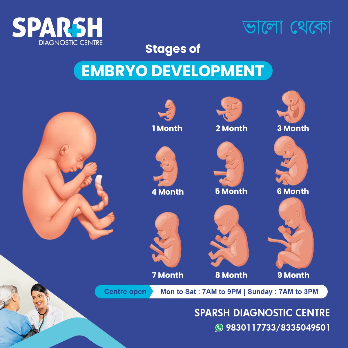
The stages of embryo development represent one of the most remarkable processes in human biology. From fertilization and the formation of a single-cell zygote, the embryo progresses through cleavage, morula and blastocyst formation, implantation, gastrulation, neurulation and organ development.
By around eight weeks after fertilization, the embryo has established the foundations of most major body structures and is subsequently referred to as a fetus. From there, development focuses increasingly on growth and maturation, with important changes continuing until birth and beyond.
For expectant parents, understanding these milestones can make pregnancy terminology much easier to follow. For those undergoing IVF, understanding the journey from zygote to blastocyst can also make laboratory updates and embryo grading easier to understand.
Most importantly, developmental timelines are guides rather than rigid schedules. Small variations in timing are common, and an individual embryo’s development should always be interpreted in the context of the pregnancy or fertility treatment by a qualified healthcare professional.
How Sparsh Diagnostic Centre Supports Pregnancy Care
At Sparsh Diagnostic Centre, we offer a full suite of prenatal diagnostic services to track your baby’s growth and ensure your health throughout pregnancy:
- Fetal Echocardiography
Fetal Well-being Assessments
Blood Investigations and Hormonal Panels
Our expert radiologists and gynaecologists provide personalized care backed by modern equipment and years of experience.
📍 Centre Timings:
Mon to Sat – 7 AM to 9 PM
Sunday – 7 AM to 3 PM
📞 Contact Us: 9830117733 / 8335049501
Embryo development is a breathtakingly intricate process that reflects the miracle of life. From a tiny cell to a full-term baby, each stage plays a crucial role in forming a healthy, thriving child. By staying informed and choosing reliable diagnostic care, parents can ensure the safest possible journey for their baby.
If you’re expecting or planning to conceive, let Sparsh Diagnostic Centre be your trusted partner in this beautiful phase of life.
#BhaloTheko
Disclaimer:
No content on this site, regardless of date, should ever be used as a substitute for direct medical advice from your doctor or other qualified clinician.