
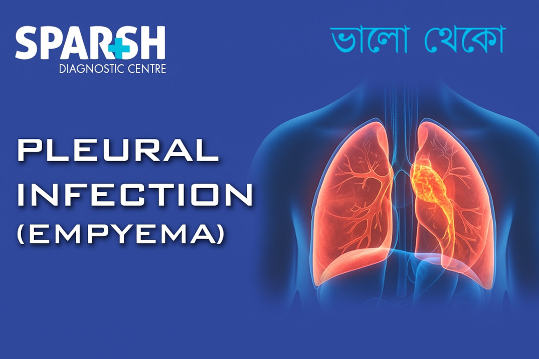
The human lungs are delicate organs protected by a double-layered membrane called the pleura. When an infection develops between these layers and pus accumulates, the condition is known as pleural infection or empyema.
Empyema is often a complication of pneumonia but may also result from chest surgery, trauma, or lung abscesses. If not treated promptly, it can cause serious breathing problems and potentially life-threatening complications.
This article provides a comprehensive overview of pleural infections — from causes and risk factors to diagnosis, treatment, and prevention — with expert insights into how diagnostic imaging and lab tests help in early detection.
What Is Pleural Infection (Empyema)?
A pleural infection occurs when bacteria, fungi, or other pathogens invade the pleural space — the narrow cavity between the lungs and the chest wall. Normally, this space contains a small amount of lubricating fluid that allows smooth lung movement during breathing.
When infection develops, pus, inflammatory cells, and bacteria accumulate in this cavity, leading to empyema. The buildup of pus can restrict lung expansion, causing severe chest pain and breathing difficulty.
Empyema typically progresses through three stages:
Exudative Stage: The pleural space fills with thin, infected fluid.
Fibrinopurulent Stage: The pus thickens, and fibrous strands form, compartmentalizing the infection.
Organizing Stage: Scar tissue develops, trapping the lung and reducing its ability to expand (known as trapped lung).
Causes of Pleural Infection (Empyema)
The primary cause of empyema is bacterial infection, most often following pneumonia. However, it can also develop after thoracic surgery, chest trauma, or as a spread from nearby infections.
Common Causative Organisms
Streptococcus pneumoniae – the most common cause, especially following pneumonia.
Staphylococcus aureus – often seen in hospital-acquired infections or post-surgery.
Klebsiella pneumoniae – common in people with diabetes or weakened immunity.
Anaerobic bacteria – frequently found in infections arising from aspiration (inhaling stomach contents).
Mycobacterium tuberculosis – responsible for tuberculous empyema, particularly in developing countries like India.
Risk Factors
Certain individuals are more prone to developing pleural infections due to underlying health or lifestyle conditions.
Major Risk Factors Include:
Weakened immune system (e.g., HIV/AIDS, chemotherapy, long-term steroids)
Recent chest or lung surgery
Alcohol abuse or malnutrition
Aspiration of foreign material (common in elderly or neurologically impaired individuals)
Symptoms of Pleural Infection
The symptoms of empyema can develop gradually or suddenly, depending on the cause and stage of infection. They are often similar to pneumonia but tend to persist longer and worsen over time.
Common Symptoms Include:
Chest pain that worsens with deep breathing or coughing
Persistent cough (sometimes producing pus-filled sputum)
Loss of appetite and weight loss
If the empyema becomes advanced, patients may also experience:
Bluish skin (cyanosis) due to poor oxygenation
Confusion or altered mental state (especially in elderly patients)
Diagnosis of Pleural Infection (Empyema)
Timely and accurate diagnosis is crucial to prevent complications like lung scarring or sepsis. Diagnosis involves a combination of clinical evaluation, imaging, and laboratory tests.
1. Medical History and Physical Examination
A doctor will first evaluate symptoms, medical history, and potential risk factors. During a physical examination, decreased breath sounds or dullness on chest percussion may indicate fluid accumulation.
2. Imaging Tests
Imaging helps confirm fluid buildup and assess its nature.
Chest X-ray: Shows pleural fluid collection or lung collapse.
Ultrasound: Helps determine whether the fluid is free-flowing or loculated (compartmentalized).
CT Scan (Computed Tomography): Provides a detailed view of the pleural cavity, revealing the extent of infection and guiding drainage procedures.
At Sparsh Diagnostic Centre, advanced imaging services assist in accurate detection and management planning.
3. Pleural Fluid Analysis (Thoracentesis)
A sample of pleural fluid is extracted using a fine needle and analyzed for:
Appearance (clear, cloudy, or pus-like)
Cell count and protein levels
Bacterial cultures and sensitivity
Glucose and pH levels
Low glucose and pH levels typically indicate infection.
4. Blood Tests
Complete blood count (CBC): Reveals elevated white blood cell count.
Blood cultures: Identify bacteria causing infection.
C-reactive protein (CRP) or ESR: Indicate inflammation levels.
Treatment of Pleural Infection (Empyema)
The goal of treatment is to eliminate infection, drain pus, and restore normal lung function. Treatment usually involves a combination of antibiotics, drainage, and sometimes surgery.
1. Antibiotic Therapy
Broad-spectrum antibiotics are started immediately and later adjusted based on culture results. Common antibiotics include:
Ceftriaxone or cefotaxime (for community-acquired infections)
Vancomycin or linezolid (for MRSA infections)
Metronidazole or clindamycin (for anaerobic bacteria)
The duration of therapy may last 2–6 weeks, depending on the severity of infection.
2. Drainage of Pus
To prevent lung compression and promote recovery, pus must be drained from the pleural space.
Needle Aspiration: Used for small, free-flowing effusions.
Chest Tube Drainage (Tube Thoracostomy): A tube is inserted into the pleural cavity to continuously drain the infected fluid.
Image-guided Drainage: Ultrasound or CT guidance ensures precise placement of the drainage tube.
3. Intrapleural Fibrinolytic Therapy
In some cases, enzymes such as urokinase or alteplase are administered into the pleural space to break down thick pus and fibrous strands, improving drainage.
4. Surgical Intervention
If antibiotics and drainage fail, surgery may be necessary.
Video-Assisted Thoracoscopic Surgery (VATS): Minimally invasive procedure to remove pus and fibrous tissue.
Open Thoracotomy: Used in severe or chronic empyema cases where the lung is trapped by scar tissue.
5. Supportive Care
Adequate hydration and nutrition
Oxygen therapy if breathing is difficult
Chest physiotherapy to improve lung expansion
Complications of Pleural Infection
Untreated or poorly managed empyema can lead to serious complications such as:
Fibrosis and trapped lung (lung cannot expand properly)
Pleural thickening or scarring
Early detection and intervention are key to preventing these outcomes.
Prevention of Pleural Infection
While not all cases of empyema can be prevented, certain measures can significantly reduce risk:
Timely treatment of pneumonia and lung infections
Good oral hygiene (to prevent aspiration pneumonia)
Regular medical check-ups for people with chronic lung disease or diabetes
Prognosis
The outlook for pleural infection largely depends on how quickly it is diagnosed and treated.
With early intervention — including appropriate antibiotics and drainage — most patients recover fully.
However, delayed treatment may result in permanent lung damage or recurrent infections.
At Sparsh Diagnostic Centre, timely imaging, laboratory testing, and consultation with respiratory specialists can ensure accurate diagnosis and effective management, helping patients breathe easier and recover faster.
When to See a Doctor
You should seek immediate medical attention if you experience:
Persistent chest pain
Fever that doesn’t subside
Difficulty breathing
Unexplained weight loss or fatigue
Prompt medical evaluation can prevent complications and improve recovery outcomes.
Pleural infection (empyema) is a serious but treatable condition. It often develops as a complication of pneumonia or after chest surgery. Recognizing symptoms early, getting diagnostic imaging and lab tests done promptly, and beginning appropriate treatment can make a significant difference in recovery.
At Sparsh Diagnostic Centre, our advanced imaging and laboratory services play a vital role in detecting pleural infections early and guiding effective treatment plans.
If you or someone you know is experiencing symptoms of pleural infection, don’t delay — consult a healthcare professional immediately.
Frequently Asked Questions (FAQ)
1. What is the difference between pleural effusion and empyema?
Pleural effusion is the buildup of fluid in the pleural cavity, while empyema is when that fluid becomes infected and pus-filled.
2. Can pleural infection resolve without drainage?
Mild infections may improve with antibiotics alone, but most cases require drainage to remove the pus and prevent lung damage.
3. Is empyema contagious?
No, empyema itself is not contagious. However, the underlying lung infection (like pneumonia) may be caused by infectious bacteria.
4. How long does it take to recover from empyema?
Recovery time varies from person to person but generally ranges from 4 to 8 weeks, depending on the severity of infection and treatment method.
5. Can empyema recur after treatment?
Recurrence is uncommon if the infection is completely cleared and underlying causes (like pneumonia or lung disease) are properly treated.
6. What diagnostic tests confirm empyema?
Chest X-ray, ultrasound, CT scan, and pleural fluid analysis (via thoracentesis) are the main diagnostic tools used to confirm empyema.
7. How serious is empyema?
Empyema is a serious condition that can cause sepsis and permanent lung damage if untreated — but with prompt treatment, the prognosis is generally good.
👉 For expert diagnostic support and imaging for pleural infections, visit Sparsh Diagnostic Centre.
#BhaloTheko
Disclaimer:
No content on this site, regardless of date, should ever be used as a substitute for direct medical advice from your doctor or other qualified clinician.

![]()


[…] Pleural infection (empyema) […]
[…] Empyema: Infection spreads to the pleural cavity. […]