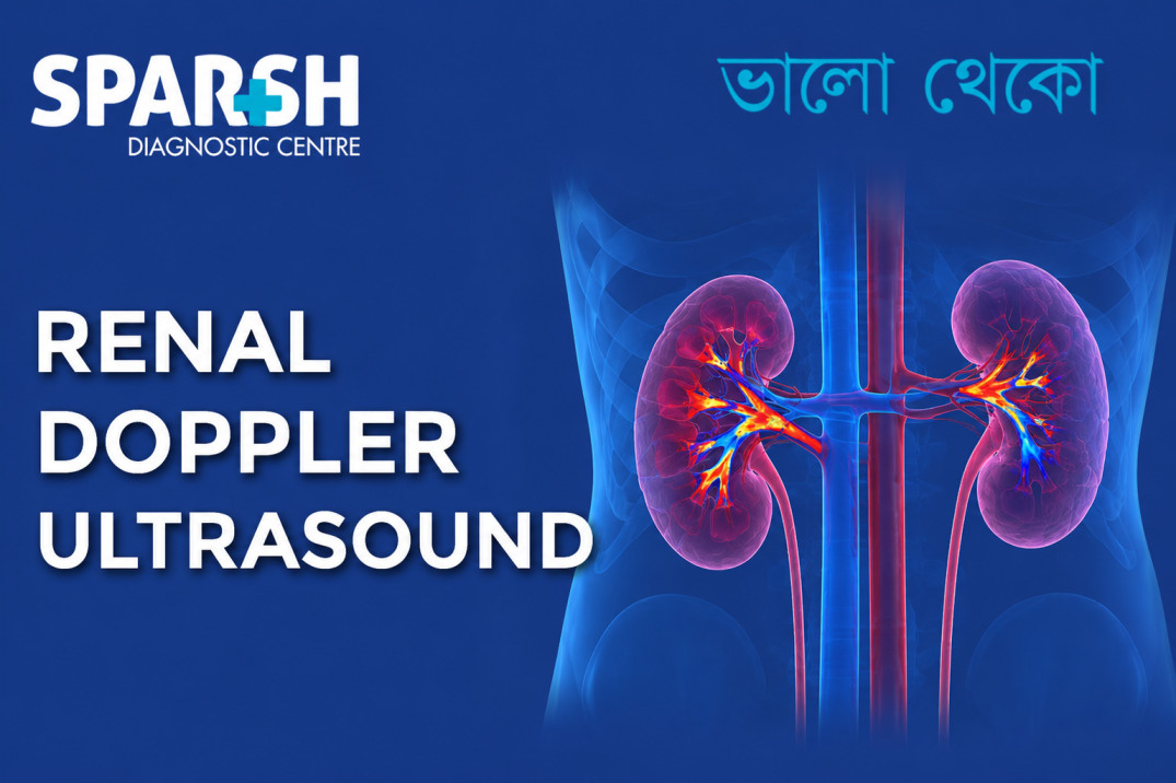
When doctors want to understand not just the structure of the kidneys but also how blood is moving through the renal arteries and within the kidneys, a renal Doppler ultrasound can provide valuable information. It is a non-invasive imaging test that combines conventional ultrasound with Doppler technology to assess blood flow.
The test is particularly useful when there is concern about renal artery stenosis, difficult-to-control high blood pressure, reduced kidney function, or problems involving a transplanted kidney. Unlike CT angiography or conventional angiography, renal Doppler does not involve ionising radiation or catheterisation.
For someone who has been advised a renal Doppler, the terminology can sound complicated. What exactly does the test look for? Do you need to fast? Can you drink water beforehand? How much does it cost? And what do the numbers on the report actually mean?
Here is a practical guide to understanding the test.
What is a renal Doppler test?
A renal Doppler test, also called a renal Doppler ultrasound, renal artery Doppler or renal color Doppler, is an ultrasound examination used to evaluate the kidneys and the blood vessels supplying them.
Regular ultrasound produces images of the kidneys, allowing the radiologist to assess their size, shape, position and general appearance. Doppler ultrasound adds information about blood flow, including the direction and speed of blood moving through the renal arteries and smaller vessels within the kidneys.
During the examination, an ultrasound probe is moved over the abdomen and sides after applying ultrasound gel to the skin. The Doppler component measures blood-flow characteristics and can help identify abnormal increases or decreases in flow.
One of the important conditions the test can help investigate is renal artery stenosis, which means narrowing of one or both arteries supplying the kidneys. Atherosclerosis is the most common cause in older adults, while fibromuscular dysplasia is more commonly seen in younger people.
A renal Doppler may be recommended for people with:
- Resistant or difficult-to-control hypertension
- Unexplained worsening kidney function
- Suspected renal artery stenosis
- Certain vascular conditions
- A history of significant cardiovascular disease
- Evaluation of a transplanted kidney
- Suspected abnormalities in renal blood flow
The test is also useful because it can provide functional information about blood flow without exposing the patient to radiation.

Why is a renal Doppler ultrasound done?
The kidneys require a continuous blood supply to perform their functions. The renal arteries carry oxygenated blood from the abdominal aorta to each kidney.
If one of these arteries becomes significantly narrowed, blood flow to the kidney may be affected. In some people, this can contribute to high blood pressure or progressive kidney dysfunction.
A renal Doppler may therefore be advised when a doctor suspects a problem with renal circulation.
It can help assess:
- The main renal arteries
- Blood-flow velocity
- Blood flow within the kidney
- Possible narrowing or obstruction
- Differences between the two kidneys
- Intrarenal blood-flow patterns
- Certain complications involving a transplanted kidney
Doppler measurements such as peak systolic velocity (PSV), renal-to-aortic velocity ratio and resistive index (RI) are among the parameters considered when assessing renal blood flow.
It is important to remember that a Doppler report should not be interpreted by looking at one number alone. The radiologist considers the Doppler measurements alongside the ultrasound appearance, symptoms, blood pressure, kidney function and other clinical information.
What happens during a renal Doppler scan?
The examination is generally straightforward.
You will usually lie on an examination table while the sonographer or radiologist applies ultrasound gel to your abdomen. A handheld transducer is then moved across different areas to obtain images of both kidneys and their blood vessels.
You may be asked to change position or briefly hold your breath. This can help reduce movement and bowel-gas interference, making it easier to visualise the renal arteries.
The Doppler component may produce sounds that resemble pulses or whooshing noises. These are simply representations of blood flow being detected by the ultrasound equipment.
There is normally no injection, incision or radiation involved.
Some patients may experience mild pressure from the probe, particularly if the examiner needs to obtain images from a deeper area, but the test is generally well tolerated.
Does a renal Doppler need fasting?
Not necessarily.
Fasting is not universally required for renal Doppler ultrasound. However, preparation instructions can differ between imaging centres. Some facilities may recommend fasting or avoiding food for several hours because food and bowel gas can sometimes make visualisation of the renal arteries more difficult.
If your diagnostic centre has specifically instructed you to fast, follow those instructions even though fasting is not an absolute requirement for every renal Doppler examination.
If you have diabetes, take regular medicines, or have another condition that makes fasting difficult, ask the centre or your doctor about the appropriate preparation before the appointment.
Can I drink water before a renal Doppler ultrasound?
Usually, yes.
Drinking water generally does not interfere with renal Doppler imaging. In fact, unlike some pelvic ultrasound examinations, a full urinary bladder is not normally the main requirement for a renal artery Doppler.
However, preparation instructions can vary depending on whether your doctor has requested a renal Doppler alone or a combination of examinations, such as an abdominal or urinary-system ultrasound.
The safest approach is to follow the instructions provided by the diagnostic centre performing the examination.
Do not deliberately dehydrate yourself before the scan unless a healthcare professional has specifically instructed you to do so.
What is the normal Doppler value for the renal artery?
There is no single number that can be called the “normal renal Doppler value.” Several measurements are evaluated.
For the main renal artery, a peak systolic velocity (PSV) of approximately 60–100 cm/s is described as a normal range in standard renal Doppler assessment. The renal-to-aortic PSV ratio is generally expected to be less than 3.5.
The resistive index (RI) is another important measurement. A commonly cited normal renal RI is approximately 0.5–0.7.
However, these numbers should not be interpreted in isolation.
For example, elevated velocity can occur with significant narrowing, but technical factors, the angle of measurement, blood-flow conditions and patient anatomy can affect Doppler readings.
Some commonly used criteria suggest that a renal artery PSV above approximately 180–200 cm/s can raise suspicion for significant stenosis, while a renal-to-aortic ratio above 3.5 is also concerning. Different laboratories and published protocols may use somewhat different thresholds.
Therefore, the radiologist’s overall impression is more important than comparing one measurement with a number found online.
How long does a renal Doppler take?
A renal Doppler ultrasound commonly takes around 30 minutes to an hour, although the exact duration varies depending on the patient’s anatomy, bowel gas, body habitus, the quality of the images and whether additional ultrasound examinations are being performed.
Some examinations may be completed relatively quickly, while others take longer when the renal arteries are difficult to visualise.
After the examination, the gel is wiped away and you can generally return to your normal activities immediately.
What are the symptoms of a blocked renal artery?
A blocked or severely narrowed renal artery does not always cause obvious symptoms.
In fact, renal artery stenosis may sometimes be discovered while investigating another medical problem.
When it does cause problems, possible signs include:
- High blood pressure that is difficult to control
- Sudden or worsening hypertension
- Declining kidney function
- Unexplained kidney dysfunction
- Episodes of fluid accumulation in the lungs or sudden breathlessness in severe cases
- Worsening blood pressure despite several medicines
Severe bilateral renal artery disease or disease affecting the artery supplying a solitary functioning kidney can be particularly important because kidney function may deteriorate significantly.
Renal artery stenosis can also cause renovascular hypertension, because reduced blood flow to the kidney can activate hormonal mechanisms that raise blood pressure.
A blocked renal artery is a medical condition that requires proper evaluation. Symptoms alone cannot confirm the diagnosis. Imaging such as Doppler ultrasound, CT angiography or MR angiography may be considered depending on the clinical situation.
At what age is renal artery stenosis most common?
The answer depends on the underlying cause.
Atherosclerotic renal artery stenosis is much more common in older adults and is associated with other vascular risk factors such as diabetes, high cholesterol, hypertension, smoking and cardiovascular disease. It is particularly encountered in people over approximately 55 years of age.
On the other hand, fibromuscular dysplasia (FMD) tends to affect younger people, particularly women. It is a non-atherosclerotic condition that can affect the renal arteries and produce renovascular hypertension.
So there is no single age at which all renal artery stenosis occurs.
In an older person with vascular risk factors, atherosclerotic disease is generally the more likely cause. In a younger person, particularly a young woman with unexplained hypertension, fibromuscular dysplasia may need to be considered.
How can I keep my renal arteries healthy?
The same habits that protect your heart and other blood vessels also help protect the renal arteries.
Control your blood pressure
High blood pressure can damage blood vessels over time. Regular monitoring and taking prescribed medicines consistently are important.
Keep cholesterol under control
High LDL cholesterol contributes to atherosclerosis, which can affect arteries throughout the body, including the renal arteries.
Do not smoke
Smoking accelerates vascular disease and increases cardiovascular risk. Stopping smoking is one of the most important steps for protecting blood vessels.
Manage diabetes
Persistently high blood glucose can damage blood vessels and increase cardiovascular risk. Good diabetes control is therefore important for long-term kidney and vascular health.
Maintain a healthy weight
A healthy body weight, combined with regular physical activity, can help reduce cardiovascular risk factors.
Stay physically active
Regular activity supports cardiovascular health and helps with blood pressure, cholesterol, weight and blood sugar management.
Eat a heart-healthy diet
A diet rich in vegetables, fruits, whole grains, legumes and appropriate sources of protein, while limiting excessive salt, highly processed foods and unhealthy fats, can support overall cardiovascular health.
People with existing kidney disease may need individual dietary advice because their requirements for protein, potassium, phosphorus and fluids can differ.
Attend regular health checks
High blood pressure, diabetes and abnormal cholesterol often cause no symptoms. Regular health checks can identify these problems early.
For people who already have atherosclerotic renal artery disease, medical management commonly includes appropriate blood-pressure control, diabetes management, lipid-lowering treatment, smoking cessation and lifestyle measures.
What is the first-line treatment for renal artery stenosis?
There is an important distinction here: the first-line treatment depends on the cause and severity of the stenosis.
For many people with atherosclerotic renal artery stenosis, initial management focuses on medical treatment and controlling cardiovascular risk factors. This can include blood-pressure management, cholesterol-lowering treatment, diabetes control, smoking cessation and lifestyle changes. Not every person with renal artery narrowing needs an angioplasty or stent.
Revascularisation may be considered in selected patients, particularly those with severe disease accompanied by high-risk features such as resistant hypertension, rapidly worsening kidney function or recurrent flash pulmonary oedema. Current guidance emphasises selecting patients carefully rather than automatically treating every detected narrowing.
For fibromuscular dysplasia, percutaneous transluminal renal angioplasty is generally the preferred revascularisation approach when intervention is indicated. Stenting is usually not required in uncomplicated FMD.
The treatment decision should therefore be made by the treating physician based on the degree of narrowing, kidney function, blood pressure, symptoms, underlying cause and overall cardiovascular risk.
What can a renal Doppler ultrasound detect?
A renal Doppler can provide information about several aspects of kidney and vascular health.
It may help identify:
- Abnormal renal artery blood-flow velocities
- Findings suggestive of renal artery stenosis
- Altered intrarenal blood-flow patterns
- Differences in renal perfusion
- Abnormal resistive indices
- Vascular abnormalities affecting a transplanted kidney
- Evidence of reduced or absent blood flow in certain circumstances
However, renal Doppler is not a complete kidney health test.
A person can have a normal renal Doppler and still have kidney disease caused by diabetes, glomerular disease, infection, medication-related injury or other conditions. Blood tests such as serum creatinine and estimated glomerular filtration rate (eGFR), urine tests and other investigations may still be necessary.
Renal Doppler ultrasound versus other imaging tests
Renal Doppler has an important advantage: it is non-invasive and does not use ionising radiation.
CT angiography can provide highly detailed images of the renal arteries but involves radiation and usually iodinated contrast. MR angiography provides another way to assess the renal vasculature and may be selected in particular situations.
Conventional catheter angiography provides detailed vascular imaging but is invasive and is generally reserved for situations where an intervention may be required or when other imaging is insufficient.
Renal Doppler is often an attractive first-line or screening examination because it is relatively accessible and can assess blood flow without radiation or arterial catheterisation.
What is the cost of a renal Doppler scan?
The renal Doppler scan cost varies between diagnostic centres, locations and the exact examination requested.
In Kolkata, currently published prices for renal Doppler or renal arterial Doppler examinations show a fairly wide range. For example, online listings include prices around ₹3,000 to ₹5,000, while some Kolkata diagnostic centres list renal arterial Doppler examinations at around ₹3,900–₹5,500.
The final cost can depend on whether the examination is:
- Renal Doppler only
- Bilateral renal artery Doppler
- Renal Doppler combined with another ultrasound
- A specialised transplant-kidney Doppler
- Part of a larger diagnostic package
If you are planning to have the test at Sparsh Diagnostic Centre, Kolkata, it is best to confirm the current price directly with the centre because diagnostic charges can change.
Frequently asked questions about renal Doppler ultrasound
Is renal Doppler painful?
No. It is generally a painless examination. You may feel some pressure from the ultrasound probe while images are being obtained.
Is renal Doppler safe?
Yes. Diagnostic ultrasound does not use ionising radiation and renal Doppler is considered a non-invasive imaging technique.
Can I go to work after a renal Doppler?
Usually, yes. There is generally no recovery period, so normal activities can typically be resumed immediately after the scan.
Does a renal Doppler detect kidney stones?
A renal Doppler is primarily designed to evaluate blood flow and renal vessels. A conventional kidney ultrasound is generally more appropriate for assessing kidney stones and urinary-system abnormalities. Sometimes both examinations are performed together.
Can renal Doppler detect renal artery stenosis?
Yes. Doppler ultrasound can identify blood-flow findings suggestive of renal artery stenosis, including increased peak systolic velocity and abnormal velocity ratios. However, difficult anatomy or technical limitations can sometimes reduce the ability to visualise the renal arteries completely.
Does a normal renal Doppler mean my kidneys are completely healthy?
Not necessarily. A normal Doppler primarily indicates that the evaluated renal blood-flow findings do not show significant abnormalities. Other kidney diseases can exist even when renal blood flow is normal.
When should you speak to a doctor about renal artery disease?
A renal Doppler may be worth discussing with your doctor if you have unexplained or resistant hypertension, worsening kidney function, significant vascular disease, or other findings that raise suspicion of impaired renal blood flow.
The test is particularly useful when there is a clinical reason to suspect renovascular disease rather than as a routine screening test for everyone.
If your doctor has recommended a renal Doppler, there is usually a specific reason for checking the blood supply to your kidneys. Taking your previous reports, blood-pressure records and relevant blood-test results to the appointment can help your doctor interpret the findings in context.
The bottom line
A renal Doppler ultrasound is a useful, non-invasive way to examine blood flow through the kidneys and renal arteries. It can play an important role in investigating renal artery stenosis, difficult-to-control hypertension, kidney dysfunction and certain problems affecting transplanted kidneys.
You generally do not need extensive preparation, although some centres may recommend fasting because bowel gas can interfere with the examination. Water is usually permitted unless your diagnostic centre gives you different instructions.
Normal Doppler measurements include several parameters rather than one universal number. A renal artery PSV around 60–100 cm/s, renal-to-aortic PSV ratio below 3.5, and intrarenal RI around 0.5–0.7 are commonly cited reference values, but interpretation depends on the complete examination.
Most importantly, an abnormal Doppler value does not automatically mean that you have a blocked renal artery. The findings need to be interpreted alongside your blood pressure, kidney function, symptoms and other imaging.
If you have been advised to undergo a renal Doppler, choosing an experienced diagnostic centre with appropriate Doppler equipment and qualified radiology expertise can help ensure that the renal arteries and intrarenal blood flow are assessed properly.
Sparsh Diagnostic Centre, Kolkata provides diagnostic imaging services for patients who require ultrasound and Doppler examinations. For appointments and current test details, contact the centre directly.
Helpline: 9830117733 / 8335049501
This article is intended for general health information and should not replace advice from your doctor. Renal Doppler findings should always be interpreted by a qualified radiologist and correlated with the patient’s clinical condition.
To consult a Doctor or get full body check-up done at Sparsh Diagnostic Centre, call our helpline numbers 9830117733/ 8335049501.
#BhaloTheko
Disclaimer:
No content on this site, regardless of date, should ever be used as a substitute for direct medical advice from your doctor or other qualified clinician.