

Breathing is something we often take for granted — until it becomes difficult. One such condition that interferes with normal breathing is atelectasis, which occurs when a portion or all of a lung collapses, preventing proper oxygen exchange. It’s not a disease in itself but a complication or sign of another underlying condition, such as lung injury, infection, or blockage of the airways.
Atelectasis can affect anyone, but it’s most common among people recovering from surgery, those with lung diseases, or individuals on mechanical ventilation. Understanding the causes, symptoms, and treatment options for atelectasis can help in early detection and management.
What Is Atelectasis?
Atelectasis refers to the collapse or closure of lung tissue, leading to a reduction or loss of gas exchange in the affected area. This means that part of the lung stops filling with air properly, resulting in reduced oxygen levels in the blood.
Normally, the lungs contain millions of tiny air sacs called alveoli, where oxygen and carbon dioxide are exchanged. In atelectasis, these alveoli deflate or fill with fluid, preventing oxygen from reaching the bloodstream effectively.
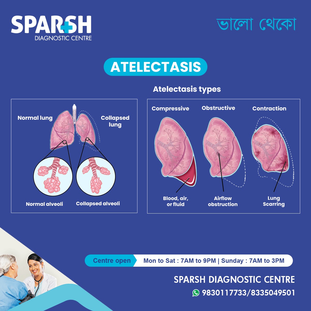
Types of Atelectasis
The infographic above highlights three major types of atelectasis — compressive, obstructive, and contraction — each with distinct underlying mechanisms.
1. Compressive Atelectasis
This occurs when something outside the lungs exerts pressure, causing part of the lung to collapse.
Common causes include:
Accumulation of fluid (pleural effusion), air (pneumothorax), or blood (hemothorax) in the pleural space
Tumors or enlarged lymph nodes pressing against the lung
Post-surgical swelling in the chest cavity
Example: A large pleural effusion due to infection or cancer can compress the lung, preventing it from expanding properly.
2. Obstructive Atelectasis
Also known as resorptive atelectasis, this type develops when there’s an airway obstruction that prevents air from reaching the alveoli. Over time, the trapped air in the lung segment gets absorbed into the bloodstream, and the alveoli collapse.
Common causes include:
Mucus plugs (especially after surgery or in people with asthma, cystic fibrosis, or COPD)
Foreign bodies blocking airways (common in children)
Tumors inside the airway (bronchial carcinoma)
Inhaled objects or thick secretions
3. Contraction (Cicatricial) Atelectasis
This type occurs when lung tissue becomes stiff or scarred, reducing its elasticity and preventing full expansion.
Common causes include:
Chronic infections or inflammatory diseases
Summary Table 🩺
| Inflammatory Disease | Mechanism | Type of Atelectasis |
|---|---|---|
| Pneumonia | Mucus/debris obstruction | Obstructive |
| Chronic Bronchitis | Airway inflammation and mucus | Obstructive |
| Asthma | Mucus plugging | Patchy/Obstructive |
| Tuberculosis | Granuloma or fibrosis | Obstructive/Fibrotic |
| Sarcoidosis | Granulomatous fibrosis | Traction |
| Hypersensitivity Pneumonitis | Alveolar inflammation | Traction |
| COPD | Chronic airway inflammation | Obstructive |
| Rheumatoid arthritis / SLE / Scleroderma | Fibrosis | Traction |
| ARDS | Alveolar inflammation | Diffuse |
| Eosinophilic Pneumonia | Inflammatory exudate | Patchy |
In this case, the lung tissue physically cannot expand properly, even when airways are clear.
Causes and Risk Factors
Atelectasis can occur due to various conditions or external factors affecting the lungs. Some of the most common causes and risk factors include:
1. Post-Surgical Complications
After surgery, especially chest or abdominal surgery, patients may breathe shallowly due to pain, anesthesia effects, or prolonged immobility. This can lead to mucus buildup and reduced airflow, causing lung collapse.
2. Airway Obstruction
Anything that blocks air passages — mucus, blood clots, foreign bodies, or tumors — can lead to obstructive atelectasis.
3. Pressure Outside the Lungs
Fluid, air, or other structures in the pleural cavity can compress lung tissue, leading to compressive atelectasis.
4. Lung Diseases
Chronic lung conditions such as asthma, COPD, or bronchiectasis can increase the risk due to recurrent infections or mucus accumulation.
5. Prolonged Bed Rest or Inactivity
When lying flat for extended periods, the lungs may not expand fully, increasing the risk of partial collapse.
6. Smoking
Smoking damages lung tissue and cilia (tiny hairs that clear mucus), promoting mucus buildup and airway blockage.
7. Mechanical Ventilation
Paradoxically, prolonged use of mechanical ventilation can cause areas of the lung to underinflate, leading to atelectasis.
Symptoms of Atelectasis
The severity of symptoms depends on how much of the lung is affected. Small areas of atelectasis may cause no noticeable signs, while larger collapses can be life-threatening.
Common symptoms include:
Rapid, shallow breathing
Coughing (sometimes dry or persistent)
Increased heart rate
In some cases, atelectasis is detected incidentally during chest X-rays taken for other reasons.
Diagnosis of Atelectasis
Timely diagnosis is essential to prevent complications like pneumonia or respiratory failure. At Sparsh Diagnostic Centre, several imaging and diagnostic tests are used to identify atelectasis and its cause.
1. Chest X-Ray
This is the first-line diagnostic test. It can show areas of lung collapse, reduced lung volume, and any associated conditions like pleural effusion or pneumothorax.
2. CT Scan (Computed Tomography)
A CT scan provides a more detailed image of the lungs, helping to pinpoint the cause (such as a tumor or mucus plug) and the exact location of the collapse.
3. Bronchoscopy
In cases of obstructive atelectasis, a bronchoscopy is performed. It allows direct visualization of the airways and helps remove mucus plugs or foreign objects.
4. Pulse Oximetry and Arterial Blood Gas (ABG)
These tests measure oxygen levels in the blood and indicate how severely gas exchange is affected.
5. Ultrasound
Useful for detecting pleural effusions or other compressive causes outside the lung.
Treatment of Atelectasis
Treatment depends on the underlying cause, extent of lung collapse, and overall health of the patient. The main goals are to re-expand the lung, remove the cause, and restore normal breathing.
1. Removing Airway Blockages
Bronchoscopy may be used to remove mucus plugs, blood clots, or foreign objects.
Chest physiotherapy helps clear secretions.
Mucolytic medications (to thin mucus) or bronchodilators may also help open airways.
2. Expanding the Collapsed Lung
Deep breathing exercises and incentive spirometry help inflate the lungs.
Positive pressure ventilation (CPAP or PEEP) can assist in keeping airways open.
Postural drainage and chest percussion may be used to mobilize mucus.
3. Treating Underlying Conditions
Antibiotics for infections (such as pneumonia or tuberculosis).
Surgery or radiation for tumors causing airway obstruction.
Drainage of pleural effusion or pneumothorax to relieve pressure on the lungs.
4. Preventing Atelectasis Post-Surgery
Early mobilization after surgery
Regular deep-breathing and coughing exercises
Use of incentive spirometers to encourage full lung expansion
Adequate pain control to enable deep breathing
Complications of Atelectasis
If left untreated, atelectasis can lead to serious complications, such as:
Permanent lung scarring or fibrosis
Prognosis and Recovery
The prognosis depends on how quickly treatment begins and the underlying cause. Small areas of atelectasis often resolve with minimal intervention, while extensive lung collapse may require hospitalization and intensive care.
With proper management, most patients recover fully, especially if the cause is addressed early. Regular follow-ups and imaging tests help ensure complete lung re-expansion.
Prevention Tips
You can reduce your risk of atelectasis with the following measures:
Stay active and avoid prolonged bed rest
Perform breathing exercises after surgery
Manage chronic respiratory conditions properly
Visit a doctor promptly if you experience persistent shortness of breath or chest pain
When to See a Doctor
Seek immediate medical help if you notice:
Sudden shortness of breath
Persistent coughing or chest discomfort
Bluish discoloration of lips or fingertips
Fever or symptoms of infection after surgery
Early diagnosis and treatment at Sparsh Diagnostic Centre can prevent serious complications and improve lung function.
FAQs About Atelectasis
1. Is atelectasis the same as a collapsed lung?
Not exactly. A collapsed lung (pneumothorax) occurs when air leaks into the space between the lung and chest wall, while atelectasis is the collapse of lung tissue itself, often due to blockage or compression.
2. Can atelectasis go away on its own?
Yes, mild atelectasis often resolves with deep breathing exercises or coughing. However, severe cases require medical intervention.
3. Is atelectasis life-threatening?
If a large portion of the lung collapses or if oxygen levels drop dangerously low, it can be life-threatening — especially in people with pre-existing lung disease.
4. How is atelectasis detected on a chest X-ray?
Radiologists look for areas of opacity (white regions) indicating collapsed lung tissue, shift of lung structures, or reduced lung volume.
5. Can children get atelectasis?
Yes. It is common in children due to mucus plugs or inhaled foreign objects blocking the airway.
6. What is the fastest way to recover from atelectasis?
Early treatment, breathing exercises, and addressing the root cause (such as removing obstructions or draining fluid) lead to faster recovery.
7. Can atelectasis recur?
Yes, particularly in people with chronic lung disease, poor airway clearance, or recurrent infections. Regular monitoring can help prevent recurrence.
Atelectasis may sound alarming, but with timely diagnosis, proper treatment, and preventive care, most people recover fully. It’s crucial to identify the cause early to prevent complications like pneumonia or respiratory failure.
If you or a loved one experiences unexplained breathing difficulties, visit Sparsh Diagnostic Centre for expert evaluation and imaging tests. Early intervention can make a significant difference in lung health and overall well-being.
#BhaloTheko
Disclaimer:
No content on this site, regardless of date, should ever be used as a substitute for direct medical advice from your doctor or other qualified clinician.

![]()


[…] Lung Collapse (Atelectasis): Due to pressure from the fluid. […]
[…] Collapsed lung (atelectasis) […]