Your heart works continuously, but you cannot directly see how well it is functioning just by checking your pulse or blood pressure. This is where a 2D echo test can be extremely useful.
A 2D echocardiogram, commonly called a 2D echo, is a non-invasive ultrasound examination that produces moving images of the heart. It allows doctors to assess the heart chambers, valves, walls, pumping action and blood flow. Unlike an X-ray, it does not use radiation.
Doctors may recommend a 2D echo when someone has symptoms such as chest discomfort, breathlessness, unexplained fatigue or a heart murmur, or when they need to monitor an already diagnosed heart condition. It can also be used as part of a broader heart-health assessment.
But a common question is: Is a 2D echo enough to detect every heart problem? The answer is no. A 2D echo is an important heart test, but different tests provide different types of information. Understanding what the test can and cannot show can help you make sense of your report.
What is a 2D echo test?
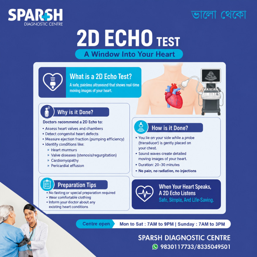
A 2D echo test is essentially an ultrasound scan of the heart. A trained professional moves an ultrasound transducer over the chest. The transducer sends high-frequency sound waves into the body and receives the returning signals. A computer converts these signals into real-time images of the beating heart.
A standard transthoracic echocardiogram, or TTE, is the most common form of echocardiography. The images are obtained from outside the chest, so there is no need to insert a probe into the body.
A 2D echo can provide information about:
- The size and shape of the heart chambers
- The thickness and movement of the heart walls
- How strongly the heart pumps
- The function of the heart valves
- The direction and speed of blood flow
- Certain congenital heart defects
- Fluid around the heart
- Some abnormalities of the heart muscle
- Certain blood clots or other structural abnormalities
One measurement doctors often look at is the ejection fraction, which helps describe how effectively the heart pumps blood.
What is a 2D echo test used for?
A 2D echo is used to evaluate the structure and function of the heart. Doctors may suggest it when they want more information than can be obtained from a physical examination or ECG alone.
Some common reasons for recommending a 2D echo include:
- Investigating chest discomfort or breathlessness
- Evaluating a heart murmur
- Checking heart valve problems
- Assessing heart pumping function
- Investigating cardiomyopathy
- Monitoring heart failure
- Looking for congenital heart abnormalities
- Checking for fluid around the heart
- Assessing changes following certain heart conditions or treatments
- Monitoring known heart disease over time
The test can also help doctors understand whether symptoms may be related to a structural or functional problem with the heart.
Importantly, a 2D echo is not only for people who already have heart disease. A doctor may recommend it because of symptoms, examination findings, risk factors or a previous abnormal test.
Why do doctors suggest a 2D echo?
Doctors suggest a 2D echo because it provides a moving picture of the heart rather than simply recording its electrical activity.
For example, if a doctor hears a heart murmur through a stethoscope, an echo can help determine whether a valve is opening or closing abnormally. If someone has breathlessness, an echo can help assess pumping function and certain structural causes.
It is also useful for monitoring known heart conditions. If a person has valve disease or reduced heart function, repeated echocardiograms can help doctors determine whether the condition is stable or changing.
In simple terms, an ECG tells doctors about the heart’s electrical activity, while an echo shows its structure, movement and blood flow.
What happens during a 2D echo?
For a standard 2D echo, you will usually lie on an examination bed, often slightly turned onto your left side.
Small electrodes may be placed on your chest to monitor your heartbeat. A gel is applied to the chest, and the ultrasound probe is gently moved over different areas to obtain images.
You may be asked to change your breathing or briefly hold your breath while certain images are recorded. The sonographer may also take measurements of the chambers, valves and blood flow.
There are no needles involved in a routine 2D echo, and the test does not expose you to X-rays. You may feel some pressure when the probe is pressed against your chest, but the procedure is generally painless.
How long does a 2D echo last?
A standard 2D echo usually takes around 30 to 40 minutes, although the exact duration can vary depending on the quality of the images required and the complexity of the examination.
Some appointments may take longer if additional views or measurements are needed.
Other forms of echocardiography take longer. For example, a stress echo involves imaging before and after exercise or medication-induced stress, while a transesophageal echocardiogram involves a probe being placed in the esophagus and requires additional preparation and recovery time.
What should I avoid before a 2D echo?
For a standard 2D echo, there is usually no special preparation. In most cases, you can eat, drink and take your usual medicines unless your doctor or diagnostic centre gives you different instructions.
It is sensible to:
- Wear comfortable clothing.
- Tell the healthcare team about your medical conditions and medicines.
- Follow any specific instructions given by your doctor.
- Avoid applying excessive lotions or oils to the chest if your centre advises against them, as they may interfere with electrode placement.
Do not stop prescribed medicines simply because you are having an echo unless your doctor specifically tells you to do so.
Preparation can be different for a stress echo or transesophageal echo. For example, some stress tests may require restrictions on caffeine, while a transesophageal echo generally requires fasting for several hours.
Is 2D echo risky?
For a standard transthoracic 2D echo, the test is considered very safe. It uses ultrasound waves rather than ionising radiation, and there are no known risks from the ultrasound itself.
The main discomfort is usually the pressure of the probe against the chest.
It is important, however, to distinguish a standard 2D echo from other types of echocardiography. A transesophageal echo involves passing a probe into the esophagus and has additional risks. Stress echocardiography can also temporarily increase the workload of the heart and therefore has different considerations.
What is the difference between an ECG and a 2D echo?
An ECG and a 2D echo are both important heart tests, but they answer different questions.
An ECG records the heart’s electrical activity through electrodes placed on the skin. It can help detect abnormal heart rhythms and certain electrical patterns associated with heart problems.
A 2D echo uses ultrasound to create moving pictures of the heart. It evaluates the heart’s chambers, valves, walls, pumping action and blood flow.
Think of it this way:
ECG = electrical activity
2D echo = structure, movement and blood flow
Neither test completely replaces the other. Depending on your symptoms, your doctor may recommend one or both.
Is 2D echo enough to detect heart problems?
Not always.
A normal 2D echo is reassuring because it may show that the heart’s structure and pumping function are normal, but it cannot rule out every possible heart problem.
For example, some rhythm disorders are better evaluated with an ECG, Holter monitor or other rhythm monitoring. Problems involving the coronary arteries may require a stress test, CT coronary angiography or coronary angiography depending on the clinical situation.
A standard echo mainly evaluates the heart’s structure, function and blood flow.
This is why doctors choose heart tests according to the person’s symptoms, medical history and risk factors rather than relying on one test for everything.
What if my 2D echo is normal?
A normal 2D echo is generally good news. It may indicate that the heart chambers, valves, pumping function and other structures assessed during the examination appear normal.
However, a normal result does not automatically mean that every possible cause of chest pain, breathlessness or other symptoms has been excluded.
For example, coronary artery narrowing may still be present even when the heart’s structure and resting pumping function look normal. Your doctor may therefore recommend additional testing if your symptoms or risk profile warrant it.
The next step depends on why the echo was ordered and what your doctor finds during the overall assessment.
Can echo confirm blockage?
A routine 2D echo does not directly show coronary artery blockages in the same way that coronary angiography or CT coronary angiography can.
An echo may show consequences of reduced blood supply, such as abnormalities in the movement of portions of the heart muscle. A stress echocardiogram can also assess how the heart responds when its workload increases and may help identify reduced blood flow to the heart muscle.
When the specific question is whether a coronary artery is narrowed or blocked, doctors may use tests such as CT coronary angiography or invasive coronary angiography. Coronary angiography is specifically used to identify narrowed or blocked coronary arteries.
Therefore, if someone asks, “Can my normal 2D echo rule out a heart blockage?” the answer is no.
What are the symptoms of heart blockage?
The phrase “heart blockage” can refer to different problems. In everyday conversation, it often means narrowing or blockage of the coronary arteries.
Possible symptoms of coronary artery disease include:
- Pressure, tightness or discomfort in the chest
- Chest discomfort that occurs with exertion
- Shortness of breath
- Pain or discomfort spreading to the arm, shoulder, back, neck or jaw
- Unusual tiredness
- Reduced exercise tolerance
Some people can have coronary artery disease with few or no obvious symptoms.
A sudden coronary blockage can cause a heart attack. Warning signs include chest pressure or pain, shortness of breath, discomfort in the arms, back, neck, jaw or stomach, sweating, nausea and lightheadedness.
What are 5 signs your heart is in danger?
There is no single list that can identify every serious heart problem, but five warning symptoms that should never be casually ignored are:
1. Chest pressure or pain: Especially pressure, squeezing, fullness or discomfort that lasts several minutes or comes and goes.
2. Breathlessness: Shortness of breath, particularly when it is new, severe or associated with chest discomfort.
3. Pain spreading to the upper body: Discomfort in the arm, shoulder, back, neck or jaw can accompany serious heart problems.
4. Sudden sweating, nausea or lightheadedness: These can occur with acute coronary syndromes, particularly when accompanied by chest discomfort.
5. Fainting or severe palpitations: Sudden fainting, near-fainting or a rapid/irregular heartbeat can sometimes indicate an important heart rhythm problem.
These symptoms do not necessarily mean that you are having a heart attack, but they deserve prompt medical assessment.
If you have sudden chest pressure or pain, severe breathlessness, fainting or symptoms suggestive of a heart attack, seek emergency medical care immediately rather than waiting for a routine 2D echo appointment.
Which echo test is best?
There is no single “best” echo test for everyone. The appropriate test depends on what the doctor needs to find out.
Standard transthoracic echocardiogram (TTE): This is the usual 2D echo performed from outside the chest. It is suitable for evaluating many structural and functional heart problems.
Doppler echocardiography: Doppler techniques assess blood flow through the heart and valves and are commonly incorporated into a comprehensive echocardiographic examination.
Stress echocardiogram: This evaluates the heart during increased workload and may be useful when doctors are investigating suspected reduced blood flow associated with exertion.
Transesophageal echocardiogram (TEE): This provides more detailed images by positioning the ultrasound probe in the esophagus, close to the heart. It may be recommended when a standard echo does not provide enough detail.
So, the “best” echo is the one that answers the clinical question your doctor is asking.
When should you consider a 2D echo?
A 2D echo may be recommended when there are symptoms or clinical findings that warrant a closer look at the heart.
It can be particularly useful when a doctor wants to assess:
- Heart pumping strength
- Heart valve function
- Heart chamber size
- Heart muscle movement
- Blood flow through the heart
- Structural abnormalities
- Fluid around the heart
- Changes associated with known heart disease
The decision should be based on an individual’s symptoms, examination and medical history rather than simply getting an echo as a routine test without a reason.
Finding reliable echocardiography services in Kolkata
If you are searching online for Best Echocardiography in Kolkata, look beyond the name of the test or the advertised price.
A good diagnostic centre should have appropriate echocardiography equipment, trained professionals, proper patient preparation, careful image acquisition and qualified interpretation of the findings.
At Sparsh Diagnostic Centre, 2D echo services are designed to provide a convenient, non-invasive assessment of heart structure and function. Patients should always discuss their symptoms and medical history with their doctor so the appropriate cardiac test can be selected.
A 2D echo can provide valuable information, but it should be interpreted as part of the bigger clinical picture.
Frequently asked questions about 2D echo
What is a 2D echo test used for?
A 2D echo is used to examine the heart’s chambers, valves, walls, pumping function and blood flow. Doctors may recommend it to investigate symptoms, assess heart murmurs, evaluate valve disease, monitor heart failure or cardiomyopathy, and look for structural heart abnormalities.
Is 2D echo enough to detect heart problems?
No. A 2D echo detects many structural and functional heart problems, but it cannot detect every heart condition. Rhythm disorders, coronary artery disease and some other problems may require ECG monitoring, stress testing, CT coronary angiography or other investigations.
What if my 2D echo is normal?
A normal echo is reassuring, but it does not rule out every heart problem. If symptoms continue, your doctor may recommend additional tests depending on the suspected cause.
What is the difference between an ECG and a 2D echo?
An ECG records the heart’s electrical activity, while a 2D echo uses ultrasound to show the heart’s structure, movement, pumping function and blood flow.
Is 2D echo risky?
A standard transthoracic 2D echo is considered very safe and does not use radiation. Mild pressure from the ultrasound probe may be felt on the chest. Other forms of echo, such as transesophageal or stress echocardiography, have additional considerations.
What are 5 signs your heart is in danger?
Important warning symptoms include chest pressure or pain, shortness of breath, pain spreading to the arm, back, neck or jaw, sudden sweating or nausea, and fainting or significant palpitations. Sudden or severe symptoms require urgent medical attention.
Can echo confirm blockage?
A routine 2D echo does not directly confirm coronary artery blockage. A stress echo may identify evidence of reduced blood flow during increased cardiac workload, while CT coronary angiography or coronary angiography can directly assess the coronary arteries.
Which echo test is best?
It depends on the medical question. A standard transthoracic 2D echo is appropriate for many routine assessments. Stress echo, Doppler techniques or transesophageal echocardiography may be more appropriate for specific clinical situations.
Why do doctors suggest a 2D echo?
Doctors recommend a 2D echo to assess heart structure, valve function, pumping ability and blood flow, investigate symptoms or abnormal examination findings, and monitor known heart conditions.
What should I avoid before a 2-D echo?
For a standard 2D echo, there is usually no need to fast or make major changes to your routine. You can generally eat, drink and take prescribed medicines as usual unless your doctor gives different instructions. Preparation can be different for stress or transesophageal echocardiography.
How long does a 2D echo last?
A standard echocardiogram commonly takes about 30 to 40 minutes, although the exact time can vary.
What are the symptoms of heart blockage?
Possible symptoms include chest pressure or discomfort, breathlessness, reduced exercise tolerance and discomfort spreading to the arm, shoulder, back, neck or jaw. A sudden blockage can cause a heart attack, which may also cause sweating, nausea or lightheadedness.
Final thoughts
A 2D echo test is one of the most useful non-invasive tools for understanding how the heart is structured and how well it is functioning. It can reveal important problems involving heart valves, chambers, heart muscle and pumping function, while Doppler imaging can provide valuable information about blood flow.
At the same time, it is important not to treat a 2D echo as a universal heart test. A normal result does not necessarily rule out coronary artery disease, rhythm disorders or every other cardiac condition. If symptoms suggest a problem, your doctor may combine an echo with an ECG, blood tests, stress testing, CT coronary angiography or other investigations.
Most importantly, do not wait for a routine diagnostic test if you develop sudden chest pressure or pain, severe breathlessness, fainting, cold sweating, nausea or pain spreading to the arm, back, neck or jaw. These can be warning signs of a heart attack and require immediate medical attention.
To consult a Doctor or get full body check-up done at Sparsh Diagnostic Centre, call our helpline numbers 9830117733/ 8335049501.
#BhaloTheko
Disclaimer:
No content on this site, regardless of date, should ever be used as a substitute for direct medical advice from your doctor or other qualified clinician.