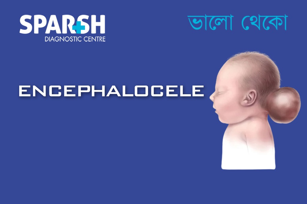
Encephalocele is a serious birth defect that affects the brain and skull of a developing baby. It occurs when part of the brain and surrounding tissues push out through an opening in the skull. This condition develops during early pregnancy when the neural tube — the structure that eventually forms the brain and spinal cord — does not close properly.
Although encephalocele can sound alarming, advances in prenatal diagnosis and surgical treatment have significantly improved outcomes for many children. Understanding the condition, its causes, and treatment options can help parents make informed decisions and seek the right medical care.
In this guide, we’ll explore everything you need to know about encephalocele, including types, symptoms, diagnosis, treatment, and long-term outlook.
What Is Encephalocele?
Encephalocele is a type of neural tube defect where brain tissue and membranes protrude through a defect in the skull. The protruding tissue forms a sac-like structure that may contain:
Brain tissue
Cerebrospinal fluid (CSF)
Protective membranes covering the brain (meninges)
The condition is usually visible at birth as a soft swelling or sac on the baby’s head. The size and location of the protrusion can vary widely, and this often influences the severity of the condition.
Encephalocele is relatively rare, occurring in approximately 1 in every 5,000 live births worldwide.

Types of Encephalocele
Encephalocele is categorized based on where the opening in the skull occurs.
1. Frontal (Anterior) Encephalocele
Frontal encephalocele occurs in the front part of the skull, typically around the forehead or nose region.
This type may cause:
Facial deformities
Nasal obstruction
Eye spacing abnormalities
Visible swelling near the forehead
In some cases, frontal encephaloceles may be surgically corrected with good cosmetic and functional outcomes.
2. Occipital (Posterior) Encephalocele
Occipital encephalocele occurs in the back of the skull, near the occipital bone.
This is the most common type of encephalocele and may involve a larger amount of brain tissue.
Possible complications include:
Developmental delays
Neurological impairment
Vision problems
The severity often depends on how much brain tissue is involved.
3. Parietal Encephalocele
This form occurs at the top of the skull. It is less common but can vary greatly in size and severity.
4. Basal Encephalocele
Basal encephaloceles occur at the base of the skull, often hidden inside the nasal cavity or sinuses. Because they are not always visible externally, diagnosis may occur later in infancy or childhood.
Symptoms may include:
Breathing difficulties
Nasal obstruction
Recurrent infections
Cerebrospinal fluid leakage
Causes of Encephalocele
The exact cause of encephalocele is not always known. However, it is linked to problems during early fetal development.
Several factors may increase the risk:
1. Neural Tube Defects
Encephalocele belongs to a group of conditions called neural tube defects, which occur when the neural tube fails to close completely during early pregnancy.
2. Genetic Factors
Some cases are associated with genetic syndromes or chromosomal abnormalities.
3. Folic Acid Deficiency
Low levels of folic acid during pregnancy can increase the risk of neural tube defects.
4. Environmental Factors
Certain environmental influences may contribute, such as:
Exposure to harmful chemicals
Maternal infections
Certain medications during pregnancy
5. Maternal Health Conditions
Conditions like diabetes or obesity may slightly increase the risk.
In most cases, encephalocele occurs sporadically, meaning there is no clear inherited cause.
Symptoms of Encephalocele
The symptoms of encephalocele depend largely on:
The size of the sac
The location of the defect
The amount of brain tissue involved
Common signs include:
Visible Symptoms
A soft swelling or sac on the baby’s head
Skin-covered or membrane-covered protrusion
Abnormal head shape
Neurological Symptoms
Developmental delays
Difficulty with movement or coordination
Vision problems
Other Possible Symptoms
Feeding difficulties
Breathing problems
Not every child with encephalocele experiences severe symptoms. Some cases involve minimal brain tissue and may have better outcomes.
How Encephalocele Is Diagnosed
Early diagnosis plays a crucial role in managing encephalocele effectively.
Prenatal Diagnosis
Many cases are detected during pregnancy through routine screening tests.
Common prenatal tests include:
Ultrasound: Often detects encephalocele during the second trimester
Fetal MRI: Provides more detailed images of the brain
Maternal blood tests: Elevated alpha-fetoprotein (AFP) levels may indicate neural tube defects
Diagnosis After Birth
If the condition is not detected during pregnancy, it is usually diagnosed immediately after birth through physical examination.
Doctors may also perform:
These tests help determine the extent of brain involvement and guide treatment planning.
Treatment for Encephalocele
Treatment usually involves surgical repair, though the approach depends on the size and location of the encephalocele.
Surgical Treatment
Surgery typically aims to:
Place brain tissue back inside the skull
Remove non-functional tissue if necessary
Close the skull opening
Reconstruct the skull and surrounding structures
Surgery is often performed within the first few months of life, though timing may vary depending on the baby’s health.
Treatment for Associated Conditions
Children with encephalocele may require treatment for additional complications such as:
Hydrocephalus
If excess fluid builds up in the brain, doctors may place a ventriculoperitoneal (VP) shunt to drain the fluid.
Seizures
Medications may be prescribed to control seizures.
Developmental Support
Therapies may include:
Physical therapy
Occupational therapy
Speech therapy
Early intervention can significantly improve quality of life.
Possible Complications
Encephalocele can sometimes lead to long-term complications depending on the severity of the condition.
Potential complications include:
Hydrocephalus
Intellectual disability
Motor skill difficulties
Vision impairment
Seizure disorders
However, outcomes vary widely. Some children with small encephaloceles and minimal brain involvement may develop normally.
Prognosis and Life Expectancy
The prognosis for encephalocele varies significantly from one child to another.
Factors that influence outcome include:
Location of the encephalocele
Size of the defect
Amount of brain tissue involved
Presence of other birth defects
Children with small anterior encephaloceles generally have better outcomes than those with large occipital ones involving significant brain tissue.
With early treatment, supportive care, and rehabilitation, many children can lead meaningful and fulfilling lives.
Can Encephalocele Be Prevented?
While not all cases can be prevented, certain steps may reduce the risk of neural tube defects.
1. Adequate Folic Acid Intake
Women planning pregnancy should take 400–800 micrograms of folic acid daily.
2. Early Prenatal Care
Regular prenatal visits allow doctors to monitor fetal development.
3. Healthy Pregnancy Lifestyle
Avoid alcohol and harmful substances
Manage chronic health conditions
Maintain balanced nutrition
These steps support healthy fetal development.
Living With Encephalocele
Raising a child with encephalocele can bring emotional and medical challenges, but many families find support through specialized healthcare teams.
Parents may work with:
Pediatric neurologists
Neurosurgeons
Developmental specialists
Physical therapists
Support groups and counseling can also help families navigate the journey.
With proper care and early intervention, children with encephalocele can achieve developmental progress and improved quality of life.
Frequently Asked Questions (FAQs)
1. Is encephalocele the same as spina bifida?
No. Both are neural tube defects, but they affect different areas. Encephalocele involves the brain and skull, while spina bifida affects the spinal cord and spine.
2. Can encephalocele be detected during pregnancy?
Yes. Most cases are detected through prenatal ultrasound, often during the second trimester. Additional tests like fetal MRI may provide more detailed information.
3. Is encephalocele life-threatening?
In severe cases, it can be life-threatening, particularly if large portions of brain tissue are involved. However, milder cases may have much better outcomes after surgery.
4. What causes encephalocele in babies?
The condition occurs when the neural tube does not close completely during early pregnancy. Genetic factors, folic acid deficiency, and environmental influences may contribute.
5. Can babies with encephalocele live normal lives?
Some children with smaller defects and successful surgery can develop normally. Others may need ongoing medical care and developmental support.
6. Is encephalocele hereditary?
Most cases are not inherited. However, certain genetic conditions may increase the risk.
7. How common is encephalocele?
Encephalocele is rare and occurs in roughly 1 in 5,000 births worldwide.
To consult a Pediatrician at Sparsh Diagnostic Centre, call our helpline numbers 9830117733/ 8335049501.
#BhaloTheko
Disclaimer:
No content on this site, regardless of date, should ever be used as a substitute for direct medical advice from your doctor or other qualified clinician.
![]()