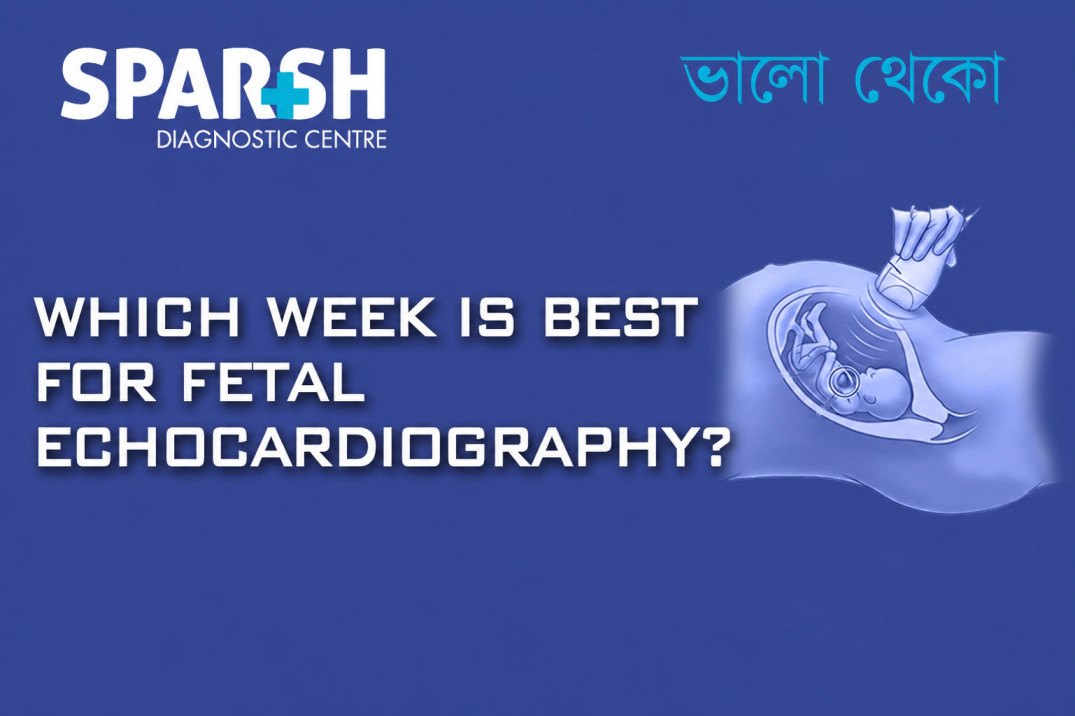
Pregnancy is filled with milestones, from hearing your baby’s heartbeat for the first time to seeing tiny movements during an ultrasound. Among the many prenatal tests available today, fetal echocardiography is one of the most important examinations for assessing the health of a baby’s heart before birth.
Many expectant parents wonder: Which week is best for fetal echocardiography? The timing of this specialized scan plays a crucial role in obtaining accurate results and ensuring early detection of potential heart abnormalities.
In this comprehensive guide, we’ll explain what fetal echocardiography is, why it is performed, the ideal time during pregnancy to undergo the test, and what parents can expect during the procedure.
What Is Fetal Echocardiography?
Fetal echocardiography is a specialized ultrasound examination that evaluates the structure and function of a baby’s heart while still in the womb. Unlike a routine pregnancy ultrasound, this test focuses exclusively on the fetal heart.
Using high-frequency sound waves, fetal echocardiography provides detailed images of the:
- Heart chambers
- Heart valves
- Blood vessels
- Blood flow patterns
- Heart rhythm and function
The examination helps doctors identify congenital heart defects and other cardiac abnormalities before birth.
Why Is Fetal Echocardiography Important?
Congenital heart defects are among the most common birth defects worldwide. Early diagnosis allows healthcare providers and parents to prepare for appropriate medical care after delivery.
Benefits of fetal echocardiography include:
- Early detection of heart defects
- Better pregnancy management
- Improved delivery planning
- Access to specialized neonatal care
- Enhanced treatment outcomes
- Reduced complications after birth
In many cases, identifying a heart condition before birth significantly improves the baby’s prognosis and quality of life.
Which Week Is Best for Fetal Echocardiography?
The ideal time for fetal echocardiography is between 18 and 24 weeks of pregnancy, with many specialists considering 20 to 22 weeks the optimal window.
During this period:
- The fetal heart is sufficiently developed.
- Cardiac structures are large enough to visualize clearly.
- Blood flow patterns can be accurately assessed.
- Most congenital heart defects can be detected.
This timing offers the best balance between fetal development and image quality, allowing specialists to conduct a thorough evaluation.
Why 20 to 22 Weeks Is Considered Optimal
At around 20 to 22 weeks:
- The baby’s heart anatomy is well-formed.
- Ultrasound imaging provides detailed visualization.
- The fetus is usually positioned favorably for examination.
- Diagnostic accuracy is highest.
If abnormalities are identified, there is adequate time for additional testing, counseling, and delivery planning.
Can Fetal Echocardiography Be Performed Earlier?
Yes, fetal echocardiography can sometimes be performed as early as 12 to 16 weeks in high-risk pregnancies.
Early fetal echocardiography may be recommended when:
- There is a strong family history of congenital heart disease.
- Previous pregnancies were affected by heart defects.
- Genetic abnormalities are suspected.
- Certain maternal medical conditions are present.
However, because the fetal heart is still very small during the first trimester, some abnormalities may not be visible. Therefore, an early scan is often followed by a detailed examination later in pregnancy.
Can Fetal Echocardiography Be Performed Later?
Yes, fetal echocardiography can also be performed during the third trimester if:
- New concerns arise during routine ultrasounds.
- Abnormal heart rhythms are detected.
- Maternal health conditions develop later in pregnancy.
- Follow-up assessments are required.
Although later examinations remain valuable, imaging quality may be somewhat limited due to fetal position, reduced amniotic fluid, or increasing fetal size.
Who Should Undergo Fetal Echocardiography?
Not every pregnant woman requires a fetal echocardiogram. However, doctors may recommend it for women with specific risk factors.
Maternal Risk Factors
A fetal echocardiogram may be advised if the mother has:
- Diabetes
- Lupus
- Phenylketonuria (PKU)
- Congenital heart disease
- Autoimmune disorders
- Exposure to certain medications during pregnancy
Family History
The test may be recommended if:
- A parent has congenital heart disease.
- A sibling has a heart defect.
- There is a strong family history of cardiac abnormalities.
Pregnancy-Related Factors
Doctors may order fetal echocardiography when:
- Routine ultrasound findings appear abnormal.
- Increased nuchal translucency is observed.
- Chromosomal abnormalities are suspected.
- IVF pregnancy is involved.
- Twin-to-twin transfusion syndrome is present.
What Conditions Can Fetal Echocardiography Detect?
Fetal echocardiography can identify a wide range of congenital heart abnormalities.
Common conditions include:
Ventricular Septal Defect (VSD)
A hole between the lower chambers of the heart.
Atrial Septal Defect (ASD)
An opening between the upper chambers of the heart.
Tetralogy of Fallot
A complex congenital heart defect involving four structural abnormalities.
Transposition of the Great Arteries
A condition where the major blood vessels are connected incorrectly.
Hypoplastic Left Heart Syndrome
Underdevelopment of the left side of the heart.
Coarctation of the Aorta
Narrowing of the aorta that restricts blood flow.
Cardiac Arrhythmias
Abnormal fetal heart rhythms that may require monitoring or treatment.
What Happens During the Procedure?
Many parents feel anxious before specialized prenatal testing. Fortunately, fetal echocardiography is safe, painless, and non-invasive.
During the procedure:
- The mother lies comfortably on an examination table.
- A gel is applied to the abdomen.
- An ultrasound probe is moved over the skin.
- Detailed images of the baby’s heart are obtained.
- Blood flow and heart function are evaluated.
The examination usually takes between 30 and 60 minutes, depending on the baby’s position and the complexity of the assessment.
Is Fetal Echocardiography Safe?
Yes. Fetal echocardiography uses ultrasound technology and does not involve radiation.
The procedure has been safely used for decades in prenatal medicine and is considered safe for both mother and baby when performed by trained professionals.
How Accurate Is Fetal Echocardiography?
Fetal echocardiography is highly accurate when performed by experienced fetal cardiologists and sonographers.
Detection rates for major congenital heart defects are often very high, particularly when the scan is conducted during the recommended 18-to-24-week period.
However, some minor abnormalities may not become apparent until after birth.
What If an Abnormality Is Found?
Receiving unexpected results can be emotionally challenging. However, prenatal diagnosis provides significant advantages.
If a heart abnormality is identified:
- Additional imaging may be recommended.
- Genetic counseling may be offered.
- Pediatric cardiologists become involved in care planning.
- Delivery arrangements can be optimized.
- Treatment options can be discussed before birth.
Many babies diagnosed prenatally go on to receive successful treatment and lead healthy lives.
Preparing for a Fetal Echocardiography Appointment
Preparation is generally simple.
Helpful tips include:
- Wear comfortable clothing.
- Carry previous ultrasound reports.
- Bring medical records if requested.
- Arrive early for registration.
- Stay hydrated unless instructed otherwise.
Some clinics may advise specific preparation guidelines depending on the stage of pregnancy.
Choosing the Right Center for Fetal Echocardiography
The accuracy of fetal echocardiography depends greatly on the expertise of the healthcare team and the quality of imaging equipment.
When selecting a center, consider:
- Experienced fetal cardiologists
- Advanced ultrasound technology
- Comprehensive prenatal care services
- Access to pediatric cardiology specialists
- Positive patient reviews
Parents searching online for “Fetal Echocardiography Near Me” should prioritize accredited diagnostic centers with expertise in fetal cardiac imaging.
If you are looking for Fetal Echocardiography Kolkata, choose a facility that offers specialized fetal medicine services and detailed prenatal cardiac evaluations.
Many expecting parents seeking the Best Fetal Echocardiography in Kolkata prefer centers equipped with high-resolution imaging systems and multidisciplinary fetal care teams.
Importance of Early Detection
One of the greatest advantages of fetal echocardiography is early diagnosis.
Early detection allows:
- Better monitoring throughout pregnancy
- Timely medical interventions
- Improved neonatal outcomes
- Reduced emergency complications after birth
- Coordinated care among specialists
For families at increased risk, fetal echocardiography offers valuable reassurance and preparedness.
Final Thoughts
So, which week is best for fetal echocardiography?
For most pregnancies, the ideal time is between 18 and 24 weeks, with 20 to 22 weeks considered the optimal period for obtaining detailed images of the baby’s heart. This timeframe allows specialists to evaluate cardiac anatomy accurately and identify most congenital heart abnormalities.
Whether recommended due to risk factors or as part of advanced prenatal care, fetal echocardiography provides critical information that can significantly improve outcomes for both mother and baby.
If your doctor recommends the test, scheduling it during the recommended window ensures the most accurate assessment possible. Families seeking Fetal Echocardiography Kolkata, the Best Fetal Echocardiography in Kolkata, or searching online for Fetal Echocardiography Near Me should consult experienced fetal medicine specialists for comprehensive prenatal cardiac evaluation.
Frequently Asked Questions (FAQs)
1. What is the best week for fetal echocardiography?
The best time is generally between 18 and 24 weeks of pregnancy, with 20 to 22 weeks considered ideal.
2. Is fetal echocardiography different from a regular pregnancy ultrasound?
Yes. Fetal echocardiography focuses specifically on the baby’s heart, while routine ultrasounds assess overall fetal growth and development.
3. How long does a fetal echocardiogram take?
The examination usually takes 30 to 60 minutes.
4. Is fetal echocardiography painful?
No. The procedure is completely non-invasive and painless.
5. Can fetal echocardiography detect all heart defects?
It can detect most major congenital heart defects, though some minor abnormalities may not be visible before birth.
6. Who needs fetal echocardiography?
Women with diabetes, congenital heart disease, abnormal ultrasound findings, family history of heart defects, or high-risk pregnancies may be advised to undergo the test.
7. Is fetal echocardiography safe for my baby?
Yes. It uses ultrasound technology and does not involve radiation exposure.
8. What happens if a heart defect is detected?
Your healthcare team may recommend additional testing, specialist consultations, and personalized pregnancy and delivery planning.
📞 Call us on +91 9830117733 or visit https://www.sparshdiagnostica.com to schedule your appointment today.
#BhaloTheko
Disclaimer:
No content on this site, regardless of date, should ever be used as a substitute for direct medical advice from your doctor or other qualified clinician.

![]()