
Pregnancy is a remarkable journey, filled with anticipation and profound changes in both the mother’s body and the developing baby. One of the most essential tools in prenatal care is the ultrasound scan, a safe and non-invasive imaging method that uses sound waves to create pictures of the baby inside the womb. From confirming pregnancy to monitoring fetal growth and identifying potential complications, ultrasound scans play a pivotal role across all three trimesters.
In this comprehensive blog, we explore what each trimester ultrasound reveals, the recommended scan schedules, and how they contribute to a healthy and informed pregnancy experience.
What is an Ultrasound Scan in Pregnancy?
Ultrasound, also known as sonography or USG (ultrasonography), is a diagnostic technique that uses high-frequency sound waves to create images of the internal structures of the body. During pregnancy, it allows healthcare providers to view the developing fetus, placenta, amniotic fluid, and uterus in real time.
Ultrasounds are considered very safe for both the mother and the baby. They are painless, do not use radiation, and can be performed at various points during the pregnancy to gather different types of information.
First Trimester Ultrasound (0–13 Weeks)
1. Dating and Viability Scan (6–9 weeks)
Purpose:
Confirms pregnancy
Detects fetal heartbeat
Estimates gestational age
Checks for multiple pregnancies
Assesses the uterus and ovaries
What it reveals:
This scan helps determine if the pregnancy is viable by checking for the presence of a fetal heartbeat. It is also used to date the pregnancy, which is crucial for establishing an accurate due date. If you’re carrying more than one baby, this scan will usually identify it.
Why it matters:
Knowing the exact gestational age is essential for planning further prenatal care. Early detection of multiple gestation or ectopic pregnancy can guide timely interventions.
2. Nuchal Translucency Scan (11–13 weeks)
Purpose:
Assesses risk of chromosomal abnormalities such as Down syndrome
Measures the nuchal translucency (fluid behind the baby’s neck)
Can be combined with blood tests for a more accurate screening
What it reveals:
The scan measures the clear (translucent) space in the tissue at the back of the baby’s neck. Increased thickness can indicate a higher risk of certain genetic conditions. This scan is often part of the combined first-trimester screening.
Why it matters:
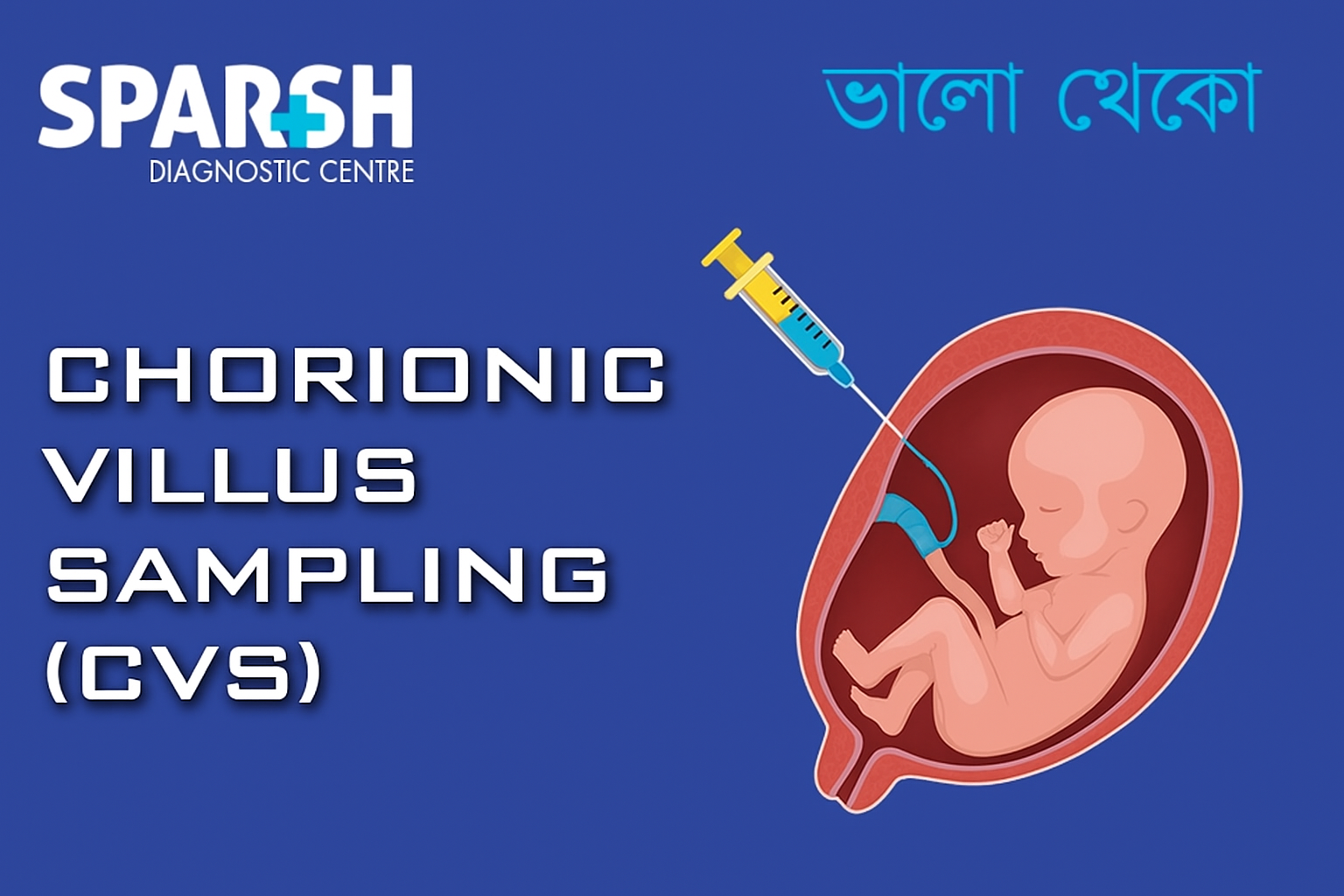
It provides an early opportunity to assess the risk of chromosomal disorders. If abnormal findings are detected, further diagnostic testing such as NIPT, CVS, or amniocentesis may be recommended.
Second Trimester Ultrasound (14–27 Weeks)
3. Anomaly Scan or Level 2 Scan (18–22 weeks)
Purpose:
Detailed assessment of fetal anatomy
Checks for congenital anomalies
Evaluates placenta position, amniotic fluid, and fetal growth
What it reveals:
This is often considered the most important scan of pregnancy. It thoroughly examines the baby’s organs, spine, brain, heart, kidneys, limbs, and facial structures. The scan can detect major birth defects and provides an overview of fetal well-being.
Why it matters:
Early detection of structural abnormalities can prepare parents and doctors for necessary interventions, specialized care, or delivery at a tertiary care center. It also assesses the placenta’s location, which is crucial in identifying conditions like placenta previa.
4. Cervical Length Scan (16–24 weeks)
Purpose:
Checks the length of the cervix
Predicts risk of preterm labor
What it reveals:
A shortened cervix during the second trimester increases the risk of premature birth. This scan is particularly important for women with a history of preterm delivery or second-trimester loss.
Why it matters:
If a short cervix is identified, doctors might prescribe medications, suggest lifestyle modifications, or consider cervical cerclage (stitching the cervix to prevent early opening).
Third Trimester Ultrasound (28–40 Weeks)
5. Growth Scan (28–32 weeks)
Purpose:
Measures fetal size and weight
Monitors fetal movements and amniotic fluid
Evaluates placenta function and position
What it reveals:
This scan checks whether the baby is growing at a normal rate. It assesses the estimated fetal weight (EFW), head circumference (HC), abdominal circumference (AC), and femur length (FL). The doctor also looks for signs of intrauterine growth restriction (IUGR) or macrosomia (excessive fetal growth).
Why it matters:
Growth scans help doctors decide if interventions are needed to ensure the baby’s health. If growth is below or above normal, further investigations or early delivery might be considered.
6. Doppler Ultrasound (30–34 weeks)
Purpose:
Measures blood flow in umbilical cord, placenta, and fetal brain
Assesses oxygen and nutrient delivery
What it reveals:
A Doppler ultrasound evaluates how well the placenta is supplying blood to the fetus. Abnormal results may indicate placental insufficiency or fetal distress.
Why it matters:
It provides crucial information in high-risk pregnancies such as gestational diabetes, hypertension, or IUGR. It helps determine if early delivery is necessary.
7. Position and Well-being Scan (36–40 weeks)
Purpose:
Checks fetal presentation (head-down or breech)
Assesses amniotic fluid levels
Monitors placental location and maturity
Confirms fetal movements and breathing
What it reveals:
This scan determines if the baby is in a favorable position for vaginal delivery. It also confirms whether the placenta has aged appropriately and checks for adequate fluid levels around the baby.
Why it matters:
If the baby is in a breech or transverse position, a cesarean delivery may be considered. This scan helps guide final decisions regarding mode of delivery and birth planning.
Ultrasound in High-Risk Pregnancies
In pregnancies with complications such as diabetes, hypertension, twins, or previous obstetric issues, ultrasounds are performed more frequently. These may include:
Serial growth scans every 2-4 weeks
Biophysical profile (BPP)
Detailed Doppler studies
Targeted scans for fetal anomalies
Regular monitoring ensures both maternal and fetal health are optimized throughout the pregnancy.
Advantages of Having Ultrasounds with a Female Radiologist
For many women, especially in conservative communities, the comfort and reassurance of being scanned by a female radiologist can make a significant difference. Sparsh Diagnostic Centre addresses this need by offering:
Evening ultrasound slots – perfect for working women
Experienced female radiologists – ensuring both comfort and competence
State-of-the-art imaging technology – for accurate results
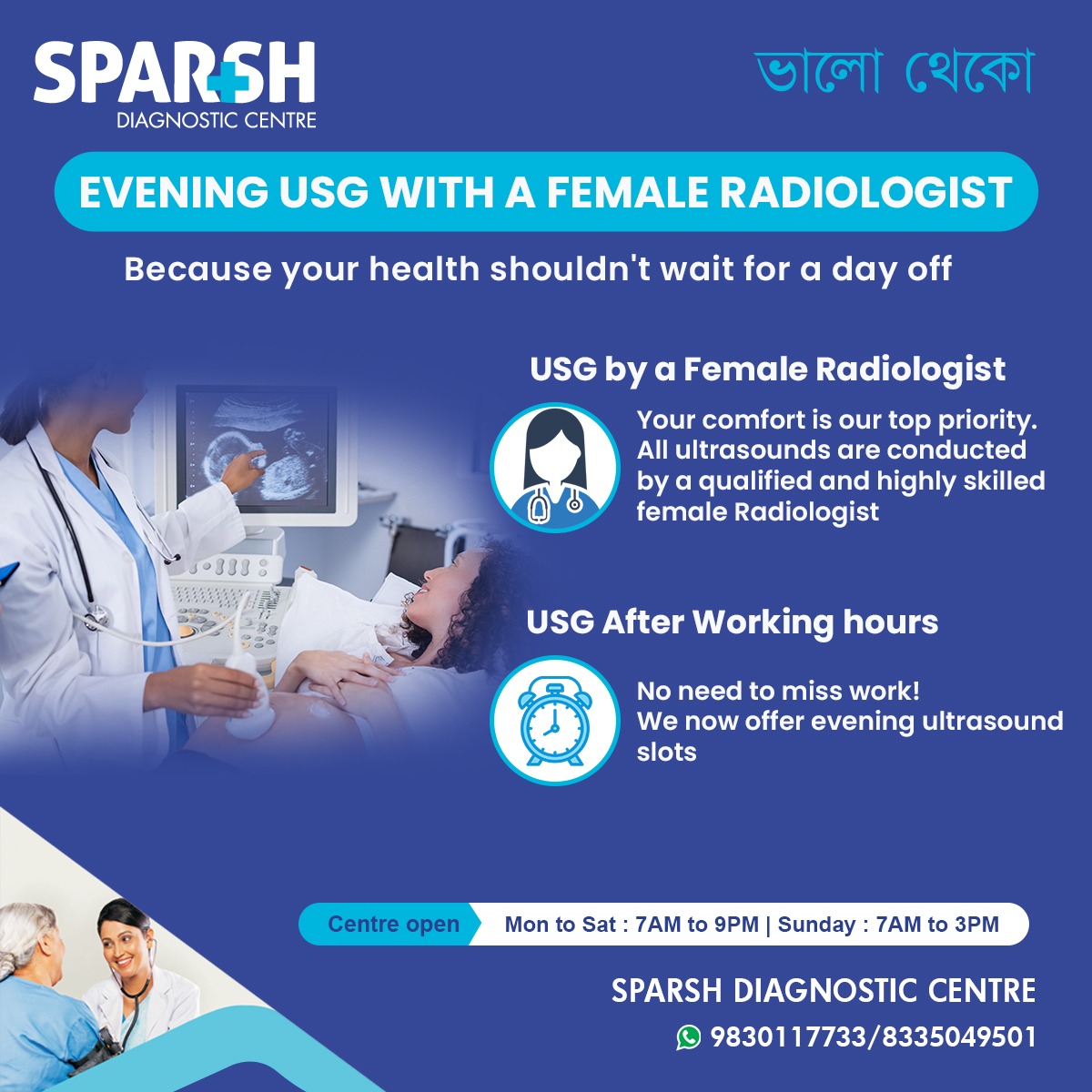
By providing ultrasounds after working hours, Sparsh ensures that women don’t have to choose between their health and their professional obligations.
Preparing for an Ultrasound Scan
Here are some tips to help you prepare for your ultrasound:
Drink water before early pregnancy scans to ensure a full bladder, which improves image quality.
Wear comfortable clothing that allows easy access to the abdominal area.
Arrive a little early to complete any paperwork and relax before your scan.
Discuss any symptoms or concerns with your doctor before the scan so the radiologist can pay special attention to specific areas.
Common Myths About Ultrasound in Pregnancy
Too many ultrasounds harm the baby – False. There is no evidence that diagnostic ultrasounds cause harm when performed correctly.
Ultrasounds always predict the exact due date – Not always. While they are accurate early in pregnancy, later estimates may vary.
Ultrasounds can detect all birth defects – Not true. While many abnormalities can be detected, some may be missed or only become apparent later.
Ultrasound always reveals the baby’s gender – Depends on the baby’s position and local regulations. In India, fetal sex determination is illegal under the PCPNDT Act.
Ultrasound scans are indispensable throughout pregnancy, providing vital insights into your baby’s health, growth, and development. Each trimester offers different revelations—from confirming the pregnancy to preparing for birth. Whether you’re undergoing a viability scan in the first trimester, a detailed anomaly scan in the second, or growth monitoring in the third, these non-invasive tests ensure you and your baby receive the best possible care.
At Sparsh Diagnostic Centre, we understand the importance of personalized, comfortable, and timely care. That’s why we offer evening ultrasound slots conducted by experienced female radiologists—because your health shouldn’t wait for a day off.
Book your ultrasound today with Sparsh Diagnostic Centre.
📞 9830117733 / 8335049501
📍 Centre open: Mon to Sat – 7 AM to 9 PM | Sunday – 7 AM to 3 PM
Because every scan tells a story—and every story matters.
#BhaloTheko
Disclaimer:
No content on this site, regardless of date, should ever be used as a substitute for direct medical advice from your doctor or other qualified clinician.

![]()

[…] of a first-trimester screening or ultrasound indicate possible […]
[…] 2. Ultrasound Imaging […]