A drooping eyelid, a smaller pupil, or reduced sweating on one side of the face may seem like minor changes, but together they can point to an important neurological condition called Horner syndrome. Although Horner syndrome itself is not a disease, it is often a sign that something has interrupted the nerve pathway connecting the brain to the eye and face.
Because this nerve pathway travels through the brain, neck, chest, and spinal cord, damage can occur in many different locations. In some cases, the cause is harmless and temporary. In others, it may indicate a stroke, carotid artery dissection, lung tumor, or spinal cord disorder that requires immediate medical attention.
The good news is that early diagnosis can identify the underlying cause, allowing doctors to begin appropriate treatment before complications develop.
This comprehensive guide explains everything you need to know about Horner syndrome, including its symptoms, causes, diagnosis, treatment, recovery, and frequently asked questions.
What is the Horner Syndrome?
Horner syndrome is a neurological disorder caused by damage to the sympathetic nerves supplying the eye and surrounding facial structures.
These sympathetic nerves control:
- Pupil size
- Eyelid position
- Sweating on the face
- Blood vessel constriction
When this nerve pathway is interrupted anywhere along its course, the affected side of the face develops a characteristic group of symptoms.
Horner syndrome can occur:
- At birth (congenital)
- During childhood
- In adults
- Suddenly after trauma
- Following surgery
- Due to neurological diseases
Importantly, Horner syndrome is not contagious and is not inherited in most cases.
Understanding the Sympathetic Nerve Pathway
To understand Horner syndrome, it helps to know how the sympathetic nerves travel.
The pathway has three neurons:
First-order neuron
Runs from the brain (hypothalamus) down through the brainstem into the spinal cord.
Damage may occur due to:
Second-order neuron
Travels from the spinal cord through the upper chest and neck.
Damage may result from:
- Lung tumors (especially Pancoast tumors)
- Neck injuries
- Thyroid surgery
- Chest surgery
Third-order neuron
Travels alongside the carotid artery into the eye.
Damage may occur because of:
- Carotid artery dissection
- Cluster headaches
- Cavernous sinus disease
Finding the exact location of damage is the most important step in treatment.
What Is the Triad of Horner Syndrome?
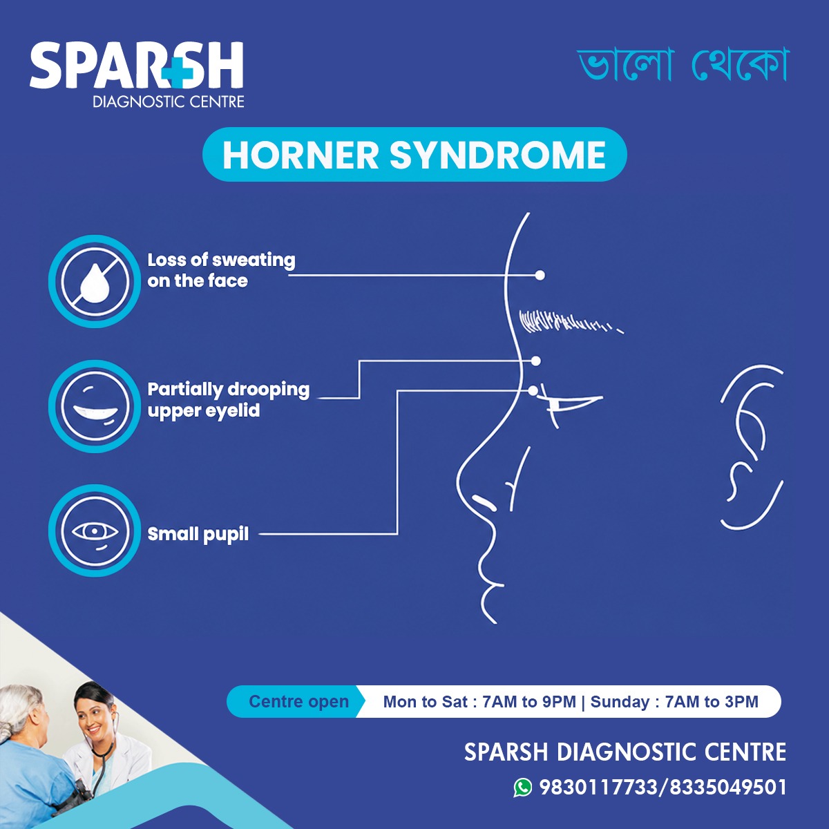
The classic triad of Horner syndrome includes:
- Ptosis (drooping upper eyelid)
- Miosis (small pupil)
- Anhidrosis (reduced sweating on the affected side of the face)
These three findings strongly suggest interruption of the sympathetic nerve pathway.
What Are the Four Signs of Horner Syndrome?
Although the classic triad is well known, doctors often describe four characteristic signs:
1. Drooping upper eyelid (Ptosis)
The eyelid droops slightly because the muscles helping lift it lose sympathetic nerve supply.
2. Small pupil (Miosis)
The affected pupil remains smaller than the normal eye and does not dilate well in darkness.
3. Reduced facial sweating (Anhidrosis)
Sweating decreases on the affected side.
4. Apparently sunken eye (Enophthalmos)
The eye may appear slightly sunken, although true displacement usually does not occur.
What Are the First Signs of Horner Syndrome?
The earliest symptoms usually include:
- One eyelid beginning to droop
- One pupil appearing smaller
- Unequal pupils becoming more obvious in dim light
- Reduced sweating on one side
- Mild facial flushing
Some patients notice changes only after comparing photographs.
Causes of Horner Syndrome
Many conditions can interrupt the sympathetic nerve pathway.
Common causes include:
Brain disorders
- Stroke
- Brain tumors
- Multiple sclerosis
- Brainstem injury
Neck disorders
- Carotid artery dissection
- Neck trauma
- Cervical spine injuries
- Neck tumors
Chest disorders
- Pancoast tumor
- Lung cancer
- Thoracic surgery
- Chest trauma
Eye-related conditions
- Cluster headaches
- Cavernous sinus disorders
- Orbital trauma
Congenital Horner Syndrome
Babies may develop Horner syndrome due to:
- Birth trauma
- Brachial plexus injury
- Neck injury during delivery
What Is Damaged in Horner Syndrome?
The damaged structure is the oculosympathetic pathway, which carries sympathetic nerve signals from the brain to the eye and face.
Depending on the location, damage may involve:
- Brainstem
- Cervical spinal cord
- Upper chest
- Sympathetic chain
- Superior cervical ganglion
- Carotid artery
- Nerves surrounding the eye
Which Artery Is Associated With Horner Syndrome?
The internal carotid artery is most closely associated with Horner syndrome.
A tear in this artery, called a carotid artery dissection, is one of the most important emergency causes.
Patients often develop:
- Sudden neck pain
- Headache
- Drooping eyelid
- Small pupil
This condition requires immediate medical evaluation because it increases stroke risk.
Which Nerve Palsy Is Associated With Horner Syndrome?
Horner syndrome itself is not a cranial nerve palsy.
However, it may occur alongside:
- Third nerve (oculomotor) palsy
- Sixth nerve palsy
- Multiple cranial nerve palsies caused by cavernous sinus disease
Doctors distinguish Horner syndrome from third nerve palsy by carefully examining pupil reactions and eyelid position.
Does Erb’s Palsy Cause Horner Syndrome?
Yes.
Erb’s palsy can occasionally be associated with Horner syndrome, particularly when birth injuries affect the lower brachial plexus and nearby sympathetic nerves.
When both occur together, doctors investigate for more extensive nerve injuries.
What Infection Causes Horner Syndrome?
Several infections can rarely damage sympathetic nerves, including:
- Tuberculosis
- Herpes zoster
- Lyme disease
- Brain abscess
- Meningitis
- Neck infections
Treating the infection often improves symptoms if nerve damage is limited.
How Do Doctors Test for Horner Syndrome?
Diagnosis begins with a thorough neurological and eye examination.
Doctors assess:
- Pupil size
- Eyelid position
- Sweating pattern
- Eye movements
- Vision
- Facial sensation
Further testing depends on suspected causes.
What Test Confirms Horner Syndrome?
Several specialized eye-drop tests help confirm the diagnosis.
Apraclonidine Test
This is currently the most commonly used confirmation test.
In Horner syndrome:
- The small pupil enlarges significantly.
- The drooping eyelid often improves.
This reversal strongly supports the diagnosis.
What Is the Hydroxyamphetamine Test for Horner Syndrome?
The hydroxyamphetamine test helps determine whether the damage involves:
- Third-order neuron
- First- or second-order neuron
How it works:
If the third-order neuron remains intact, hydroxyamphetamine stimulates norepinephrine release and the pupil dilates.
If the third-order neuron is damaged, the pupil fails to dilate.
Although useful, this test is less commonly performed today because imaging techniques have improved.
Other Tests Used for Horner Syndrome
Doctors may recommend:
MRI
To evaluate:
- Brain
- Brainstem
- Neck
- Spinal cord
CT Scan
Useful for detecting:
- Lung tumors
- Neck masses
- Chest abnormalities
CT Angiography or MR Angiography
These evaluate:
- Carotid artery dissection
- Blood vessel abnormalities
Chest Imaging
Especially important in smokers or patients with suspected lung disease.
Differential Diagnosis of Horner Syndrome
Several conditions can resemble Horner syndrome.
These include:
- Third cranial nerve palsy
- Physiological anisocoria
- Adie’s tonic pupil
- Myasthenia gravis
- Mechanical eyelid drooping
- Orbital trauma
- Chronic eye inflammation
Accurate diagnosis is essential because treatment varies widely.
What Eye Drops Are Used for Horner Syndrome?
Eye drops are mainly used for diagnosis rather than treatment.
These include:
Apraclonidine
Most commonly used today to confirm Horner syndrome.
Cocaine Eye Drops
Previously considered the gold standard but now used infrequently due to limited availability.
Hydroxyamphetamine
Used to localize the lesion within the sympathetic pathway.
These medications should only be administered under medical supervision.
What Nerve Block Causes Horner Syndrome?
Certain medical procedures involving nerve blocks can temporarily cause Horner syndrome.
Examples include:
- Stellate ganglion block
- Cervical epidural anesthesia
- Interscalene brachial plexus block
In these situations, symptoms usually resolve after the anesthetic wears off.
How Is Horner Syndrome Treated?
There is no single treatment for Horner syndrome itself.
Treatment focuses on correcting the underlying cause.
Examples include:
Stroke
- Emergency stroke treatment
- Rehabilitation
Carotid artery dissection
- Blood-thinning medication
- Specialist care
Lung tumor
- Surgery
- Chemotherapy
- Radiation therapy
Infection
- Antibiotics
- Antiviral medication
Multiple sclerosis
Can Horner Syndrome Be Reversed?
Sometimes.
Recovery depends entirely on the cause.
Possible outcomes include:
Complete recovery
Occurs when nerve compression or temporary injury resolves.
Partial recovery
Some eyelid drooping or pupil asymmetry may remain.
Permanent symptoms
If nerve fibers are permanently damaged, some signs may persist indefinitely.
How Long Can Horner Syndrome Last?
Duration varies considerably.
Temporary cases caused by:
- Local anesthesia
- Surgery
- Minor trauma
may resolve within days or weeks.
Other cases caused by nerve injury may last:
- Several months
- Years
- Permanently
Is Horner Syndrome an Emergency?
Sometimes—yes.
Seek immediate medical attention if Horner syndrome develops suddenly, especially when accompanied by:
- Severe headache
- Neck pain
- Stroke symptoms
- Vision changes
- Difficulty speaking
- Weakness
- Loss of balance
These symptoms may indicate a carotid artery dissection or stroke, both of which require urgent evaluation.
If Horner syndrome develops gradually without other concerning symptoms, it still warrants prompt medical assessment to identify the underlying cause.
What Age Does Horner Syndrome Appear?
Horner syndrome can develop at any age.
It may appear:
- At birth
- During infancy
- Childhood
- Adulthood
- Older age
The cause often varies by age group. Congenital cases are usually related to birth trauma or developmental abnormalities, while adult cases are more often linked to neurological disorders, trauma, vascular conditions, or tumors.
Living With Horner Syndrome
Most people adapt well to the physical changes caused by Horner syndrome. The biggest priority is identifying and treating the underlying condition.
Regular follow-up with a neurologist or ophthalmologist may include:
- Monitoring pupil changes
- Checking eyelid function
- Assessing vision
- Repeat imaging when necessary
If the underlying cause is successfully treated, many patients experience stabilization or improvement in symptoms.
When Should You See a Doctor?
Arrange medical evaluation if you notice:
- One pupil becoming noticeably smaller than the other
- A new drooping eyelid
- Reduced sweating on one side of the face
- Sudden changes in pupil size
- Persistent facial asymmetry
- Symptoms following neck or chest injury
Urgent evaluation is especially important if these changes occur suddenly or are accompanied by severe headache, neck pain, weakness, numbness, difficulty speaking, or vision loss.
Frequently Asked Questions (FAQs)
1. What is Horner syndrome?
Horner syndrome is a neurological condition caused by disruption of the sympathetic nerve pathway to the eye and face, resulting in a drooping eyelid, a small pupil, and reduced facial sweating on the affected side.
2. What is the triad of Horner syndrome?
The classic triad consists of ptosis (drooping eyelid), miosis (small pupil), and anhidrosis (reduced facial sweating).
3. What are the four signs of Horner’s syndrome?
The four commonly described signs are:
- Ptosis
- Miosis
- Anhidrosis
- Apparent enophthalmos (a sunken-eye appearance)
4. What is the hydroxyamphetamine test for Horner syndrome?
It is a diagnostic eye-drop test that helps determine whether the nerve damage is located in the third-order sympathetic neuron by assessing the pupil’s ability to dilate.
5. What test confirms Horner syndrome?
The apraclonidine eye-drop test is the most commonly used test to confirm Horner syndrome. Imaging studies such as MRI, CT scans, or CT angiography are then used to identify the underlying cause.
6. Which nerve palsy is associated with Horner syndrome?
Although Horner syndrome is not a cranial nerve palsy, it can occur alongside third cranial nerve (oculomotor) palsy or other cranial nerve disorders depending on the underlying condition.
7. What is a differential diagnosis of Horner’s syndrome?
Conditions that may mimic Horner syndrome include third nerve palsy, physiological anisocoria, Adie’s tonic pupil, myasthenia gravis, orbital trauma, and mechanical eyelid ptosis.
8. Does Erb’s palsy cause Horner’s syndrome?
Yes. Horner syndrome may occur with Erb’s palsy or other brachial plexus injuries when sympathetic nerve fibers are damaged, particularly after birth trauma.
9. What nerve block causes Horner’s syndrome?
Stellate ganglion blocks, cervical epidural anesthesia, and interscalene brachial plexus blocks may temporarily produce Horner syndrome.
10. Which artery is associated with Horner’s syndrome?
The internal carotid artery is most commonly associated, especially in cases of carotid artery dissection.
11. What infection causes Horner’s syndrome?
Rare causes include tuberculosis, herpes zoster, Lyme disease, meningitis, brain abscesses, and deep neck infections.
12. How do you treat Horner’s syndrome?
Treatment focuses on the underlying cause, such as managing a stroke, repairing a carotid artery dissection, treating infections, or addressing tumors. There is no specific cure for Horner syndrome itself.
13. What is damaged in Horner’s syndrome?
The oculosympathetic nerve pathway, which runs from the brain to the eye and face, is damaged.
14. What eye drops are used for Horner’s syndrome?
Apraclonidine, hydroxyamphetamine, and, less commonly, cocaine eye drops are used primarily for diagnostic purposes.
15. Is Horner’s syndrome an emergency?
It can be. Sudden-onset Horner syndrome, especially with severe headache, neck pain, or neurological symptoms, requires immediate emergency evaluation.
16. What are the first signs of Horner syndrome?
Early signs include a drooping eyelid, a smaller pupil on one side, unequal pupils that are more noticeable in dim light, and decreased facial sweating.
17. How long can Horner’s syndrome last?
It may be temporary or permanent. Recovery depends on the cause and the extent of nerve damage.
18. How do you reverse Horner’s syndrome?
There is no direct reversal. Symptoms may improve if the underlying condition is treated promptly and the nerve pathway recovers.
19. How do you test for Horner’s syndrome?
Doctors perform a detailed eye examination, assess pupil responses, use apraclonidine eye drops for confirmation, and may order MRI, CT scans, or vascular imaging to determine the underlying cause.
20. What age does Horner’s syndrome appear?
Horner syndrome can occur at any age, from newborns with birth-related injuries to older adults with neurological or vascular conditions.
21. Which nerve palsy causes Horner’s syndrome?
Horner syndrome results from damage to the sympathetic nerve pathway rather than a cranial nerve palsy, although it may coexist with third cranial nerve palsy in certain neurological disorders.
Final Thoughts
Horner syndrome is more than an eye condition—it is often a valuable clinical clue that points to an underlying neurological, vascular, or chest disorder. While the characteristic signs of a drooping eyelid, a smaller pupil, and reduced facial sweating may appear subtle, they should never be ignored. Prompt evaluation can help identify serious causes such as stroke, carotid artery dissection, or lung tumors, allowing timely treatment and improving outcomes.
If you or someone you know develops symptoms suggestive of Horner syndrome, consult an ophthalmologist or neurologist as soon as possible. Early diagnosis and appropriate investigations are key to managing both the syndrome and its underlying cause.
To consult a Doctor or get full body check-up done at Sparsh Diagnostic Centre, call our helpline numbers 9830117733/ 8335049501.
#BhaloTheko
Disclaimer:
No content on this site, regardless of date, should ever be used as a substitute for direct medical advice from your doctor or other qualified clinician.