


Pregnancy comes with a long list of scans and tests, and it is completely understandable to wonder whether every test is really necessary. One question that often comes up after the second-trimester anomaly scan is: Do you need a fetal echo if the anomaly scan is normal?
The short answer is: not necessarily.
A normal anomaly scan is reassuring and means that no obvious structural abnormality was identified during that examination. However, a fetal echocardiogram is a more detailed examination specifically designed to assess the baby’s heart. Whether you need one depends on your pregnancy history, the findings on your ultrasound scans, family history and other risk factors.
A routine anomaly scan already includes an assessment of the fetal heart, including views that screen for major heart abnormalities. A fetal echocardiogram goes further and provides a more comprehensive assessment of the heart’s structure and function.
So, having a normal anomaly scan does not automatically mean that a fetal echo is required. At the same time, a normal anomaly scan does not always mean that a fetal echo is unnecessary.
Let’s look at the difference and when a fetal echo may be recommended.
What is an anomaly scan?
The anomaly scan, also called the anatomy scan or the 20-week scan, is a detailed ultrasound examination usually performed during the second trimester. It looks at the baby’s overall anatomy and development, including the brain, face, spine, abdomen, limbs, kidneys and heart.
The fetal heart is also assessed as part of routine cardiac screening. This generally includes the four-chamber view and assessment of the outflow tracts and major vessels. A thorough cardiac screening examination improves the ability to detect congenital heart defects.
The anomaly scan is therefore much broader than a fetal echo.
It asks questions such as:
- Is the baby’s overall anatomy developing normally?
- Are the major organs formed appropriately?
- Is the baby’s growth appropriate for the gestational age?
- Does the spine appear normal?
- Are the kidneys, stomach, bladder and limbs developing normally?
- Does the fetal heart appear structurally normal on screening views?
A normal anomaly scan is good news. But it is still a screening examination rather than a guarantee that every possible condition has been ruled out.
What is a fetal echo?
A fetal echocardiogram, or fetal echo, is a specialized ultrasound examination focused specifically on the baby’s heart.
Instead of assessing the entire baby, the examination concentrates on cardiac anatomy and function. Depending on the clinical situation, the specialist may evaluate the four chambers, valves, outflow tracts, major blood vessels, heart rhythm and blood flow.
The American Heart Association describes fetal echocardiography as a detailed ultrasound that evaluates the structure and function of the developing baby’s heart. It is generally recommended when there is an increased risk of congenital heart disease.
This is why an anomaly scan and fetal echo should not be considered interchangeable tests.
Think of the anomaly scan as a broad health check of the baby’s anatomy, while a fetal echo is a specialized examination of the heart.
Is fetal echo necessary after an anomaly scan?
Not for every pregnancy.
If the anomaly scan is completely normal, there are no significant maternal or family risk factors, and the treating doctor has no reason to suspect a heart problem, a separate fetal echo may not be necessary.
However, your doctor may recommend fetal echocardiography even when the anomaly scan appears normal if there is a known risk factor for congenital heart disease.
According to current fetal cardiac screening guidance, indications can include a suspected heart abnormality, certain fetal abnormalities, increased nuchal translucency, a known or suspected genetic condition, a first-degree family history of congenital heart disease, pre-existing diabetes, certain maternal antibodies, and specific medication or environmental exposures.
This is an important distinction: the decision is based on the whole clinical picture, not just the words “normal anomaly scan.”
What if an anomaly scan is normal?
If your anomaly scan is normal, there is generally no reason to panic.
A normal scan means that the structures assessed during the examination appeared normal at that point in pregnancy. It is reassuring and is an important part of routine prenatal care.
However, ultrasound cannot detect every heart condition. Some congenital heart defects can be difficult to identify during routine screening, particularly abnormalities that may not be obvious on the four-chamber view alone. This is one reason why assessment of the outflow tracts and great vessels is important.
There is also another important point: some heart problems become apparent later in pregnancy or may be difficult to identify before birth.
Therefore, if your obstetrician recommends a fetal echo despite a normal anomaly scan, it does not necessarily mean that something is wrong. The recommendation may simply be because your pregnancy falls into a higher-risk category or because a more detailed cardiac assessment would be useful.
Does every pregnant woman get a fetal echo?
No.
Every pregnant woman does not routinely need a fetal echocardiogram.
Routine fetal cardiac screening is part of the detailed second-trimester ultrasound examination, but a comprehensive fetal echocardiogram is generally reserved for pregnancies in which there is an increased risk of congenital heart disease or a concern has been identified.
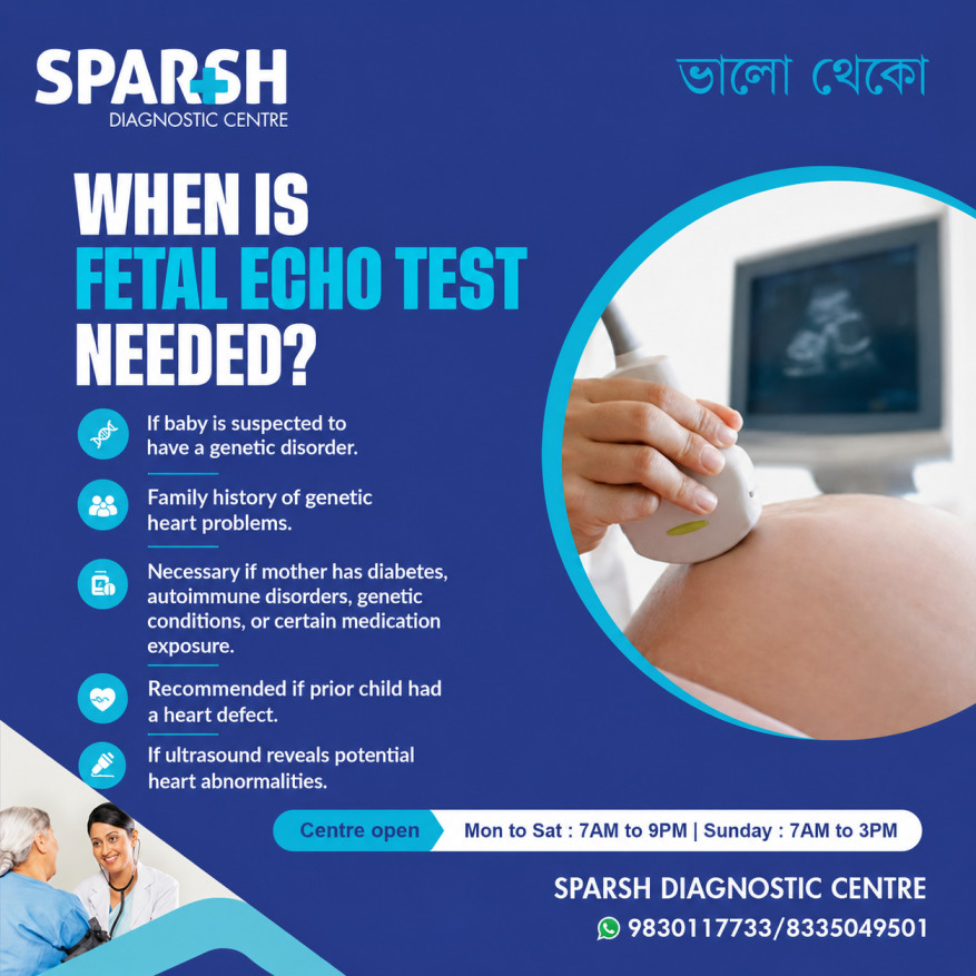
For example, fetal echo may be recommended when:
- A previous child had congenital heart disease.
- The mother or father has certain congenital heart conditions.
- There is a family history of congenital heart disease.
- The baby has a suspected heart abnormality on ultrasound.
- Another fetal abnormality has been detected that is associated with heart defects.
- The nuchal translucency measurement was significantly increased.
- A genetic abnormality is suspected or confirmed.
- The mother has pre-existing diabetes.
- Certain medications or exposures may increase the risk of congenital heart disease.
- There are specific maternal antibodies, such as anti-Ro/SSA antibodies.
- The pregnancy involves certain fetal conditions, including some monochorionic twin pregnancies.
The exact recommendation can vary depending on the clinical situation and the expertise available at the center performing the scan.
Is it necessary to do a fetal echo?
A fetal echo is necessary when there is a medical reason to take a closer look at the baby’s heart.
It is not automatically necessary simply because you are pregnant.
For example, if your anomaly scan shows a possible cardiac abnormality, your doctor may recommend a fetal echo to clarify the finding. Similarly, if you have a significant family history of congenital heart disease or pre-existing diabetes, your doctor may recommend fetal echocardiography even if the routine cardiac screening appears normal.
In other cases, your doctor may decide that the routine cardiac assessment performed during the anomaly scan is sufficient.
The best approach is therefore not to decide based solely on whether your anomaly scan was normal. Consider why the fetal echo has been suggested in the first place.
Which week is best for fetal echo?
The optimal time for a comprehensive fetal echocardiogram is generally between 18 and 22 weeks of pregnancy.
Both the American Society of Echocardiography and the American Heart Association identify this period as the usual window for fetal echocardiography.
Within this window, the exact timing can depend on the reason for the examination, fetal position, image quality, local practice and the specialist’s assessment.
The International Society of Ultrasound in Obstetrics and Gynecology also recommends cardiac screening optimally between 18 and 22 weeks. It notes that examinations performed around 20–22 weeks may be less likely to require an additional scan for completion compared with earlier examinations.
So if your doctor has recommended a fetal echo, scheduling it around this period is generally ideal.
Is a fetal echo at 23 weeks necessary?
A fetal echo at 23 weeks can still be appropriate, particularly if your doctor has identified a reason for detailed cardiac assessment.
The ideal window is usually 18–22 weeks, but that does not mean the examination suddenly becomes ineffective after 22 weeks. Many fetal cardiac structures can still be assessed satisfactorily beyond 22 weeks.
If your anomaly scan was performed around 20–22 weeks and your doctor subsequently recommends a fetal echo, having it at 23 weeks may be perfectly reasonable.
There is generally no benefit in delaying an indicated fetal echo simply because the ideal window has passed.
Is it okay to get a fetal echo at 26 weeks?
Yes, a fetal echo can still be performed at 26 weeks when clinically indicated.
The usual target is 18–22 weeks because this provides an excellent window for detailed assessment, but later examinations are still possible. Ultrasound visualization of many fetal cardiac structures remains possible beyond 22 weeks.
If you are already 26 weeks pregnant and have been advised to have a fetal echo, you should not assume that it is “too late.” Speak to your obstetrician or fetal medicine specialist and arrange the examination as advised.
The important thing is that an indicated test should not be unnecessarily postponed.
Why might a fetal echo be advised after a normal anomaly scan?
This is one of the most common sources of confusion for parents.
A normal anomaly scan can be reassuring while a fetal echo is still recommended because the two examinations have different purposes.
For example, a doctor may recommend fetal echo because:
There is a family history of congenital heart disease.
If a parent or close family member has certain congenital heart conditions, the baby’s risk may be higher than that of the general population.
The mother has pre-existing diabetes.
Pre-existing diabetes is recognized as a risk factor for congenital heart disease and is among the indications for fetal echocardiography.
The nuchal translucency was increased earlier in pregnancy.
An increased nuchal translucency measurement can be an indication for detailed cardiac assessment even if a later measurement appears normal.
The ultrasound specialist wants a closer look at the heart.
Sometimes the routine scan raises a question about the cardiac anatomy or image quality is insufficient to confidently assess all required views. A fetal echo can provide a more detailed evaluation.
There is another fetal abnormality.
Some abnormalities outside the heart are associated with congenital heart disease, so a detailed cardiac examination may be recommended.
In these situations, a fetal echo is not necessarily being ordered because your baby definitely has a heart problem. It is often being performed because the doctor wants to be particularly thorough.
What does a fetal echo check?
A fetal echo can provide a detailed assessment of several aspects of the baby’s cardiovascular system.
Depending on gestational age and the clinical indication, the specialist may examine:
- The four chambers of the heart
- The heart valves
- The ventricles and atria
- The ventricular septum
- The outflow tracts
- The major arteries
- The great vessels
- Heart rhythm
- Blood flow through the heart and vessels
- Overall cardiac function
Some congenital heart defects can be difficult to detect if only a four-chamber view is considered. Assessment of the outflow tracts and great vessels therefore adds important information to fetal cardiac screening.
Can a normal fetal echo guarantee a healthy heart?
No test can guarantee that a baby will have no health problems.
A normal fetal echocardiogram is very reassuring and means that no significant abnormality was identified during the examination. However, some cardiac problems are difficult to detect prenatally, while others may develop or become more apparent later.
The purpose of fetal echo is to improve the assessment of the baby’s heart and identify significant abnormalities before birth when possible.
If the result is normal, your doctor will generally continue your pregnancy care according to your individual circumstances.
What should you do if your doctor recommends fetal echo?
First, don’t assume that the recommendation means something is wrong.
Ask your doctor why the examination has been suggested. The reason might be something found during the ultrasound, a family history, maternal diabetes, a previous pregnancy affected by congenital heart disease, an earlier ultrasound finding, or another risk factor.
You can also ask:
- Was anything unusual seen during my anomaly scan?
- Is the fetal echo being recommended because of a risk factor?
- Does my baby’s heart look normal on the routine scan?
- When should I have the fetal echo?
- Will I need another cardiac scan later?
Understanding the reason for the test can make the process much less stressful.
Fetal echo after a normal anomaly scan: the bottom line
So, do you need a fetal echo if you have a normal anomaly scan?
Not necessarily.
A normal anomaly scan is reassuring, and many pregnant women do not need a separate fetal echocardiogram. However, fetal echo may still be recommended when there are specific risk factors or when the doctor wants a more detailed evaluation of the baby’s heart.
The usual timing for fetal echocardiography is 18–22 weeks, although it can still be performed later when clinically necessary. A fetal echo at 23 weeks or even 26 weeks may therefore still be appropriate when recommended by your doctor.
Most importantly, don’t compare your pregnancy with someone else’s. Two women can have equally normal anomaly scans but receive different recommendations because their medical histories and risk factors are different.
If you have been advised to undergo fetal echocardiography, discuss the reason with your obstetrician or fetal medicine specialist. The goal is not to add unnecessary tests, but to make sure the baby’s heart receives the appropriate level of assessment for your individual pregnancy.
Frequently asked questions
Is it necessary to do a fetal echo?
A fetal echo is not necessary for every pregnancy. It is generally recommended when there is an increased risk of congenital heart disease, a suspected abnormality on ultrasound, or another specific clinical indication.
Does every pregnant woman get a fetal echo?
No. Every pregnant woman does not routinely require a separate fetal echocardiogram. Routine second-trimester ultrasound includes fetal cardiac screening, while a comprehensive fetal echo is generally performed when there is a specific indication.
What if an anomaly scan is normal?
A normal anomaly scan is reassuring. It means that no obvious abnormality was identified during the examination. However, certain risk factors may still lead your doctor to recommend a fetal echo, even when the routine scan looks normal.
Is a fetal echo at 23 weeks necessary?
A fetal echo at 23 weeks can be appropriate when there is a clinical reason for the examination. Although 18–22 weeks is generally considered the optimal window, many cardiac structures can still be evaluated after 22 weeks.
Is fetal echo necessary after an anomaly scan?
Not automatically. If the anomaly scan is normal and there are no significant risk factors, your doctor may not recommend a separate fetal echo. However, specific maternal, fetal or family factors can make fetal echocardiography appropriate even after a normal anomaly scan.
Is it okay to get a fetal echo at 26 weeks?
Yes. Although 18–22 weeks is the usual optimal window, a fetal echo can still be performed at 26 weeks when medically indicated. If your doctor has recommended the examination, it is better to arrange it rather than assume that 26 weeks is too late.
Which week is best for fetal echo?
The usual recommended window for a comprehensive fetal echocardiogram is 18 to 22 weeks of pregnancy. Around 20–22 weeks can be particularly useful because fetal cardiac structures are generally well visualized at this stage.
Is fetal echo safe?
Fetal echocardiography uses ultrasound rather than ionizing radiation. It is considered a safe diagnostic examination when performed appropriately. The American Heart Association describes it as an ultrasound examination used to evaluate the developing baby’s heart.
Does a fetal echo replace the anomaly scan?
No. A fetal echo and anomaly scan have different purposes. The anomaly scan evaluates the baby’s overall anatomy and includes cardiac screening, while fetal echocardiography provides a specialized and more detailed examination of the heart.
What should I do if my anomaly scan is normal but my doctor recommends fetal echo?
Ask what specific risk factor or finding prompted the recommendation. A fetal echo may be advised as an additional precaution because of your medical history, family history, an earlier ultrasound finding or another recognized risk factor. A recommendation for fetal echo does not automatically mean that your baby has a heart defect.
Need a fetal echo or pregnancy ultrasound? Discuss the appropriate scan and timing with your obstetrician or fetal medicine specialist. At Sparsh Diagnostic Centre, pregnancy imaging services are available from Monday to Saturday, 7 AM to 9 PM, and Sunday, 7 AM to 3 PM. For appointments, call 9830117733 or 8335049501.
Why Choose Sparsh Diagnostic Centre for Fetal Echo?
High-resolution fetal ultrasound equipment
Trained pediatric cardiologists and fetal medicine experts
Compassionate care and clear reporting
Quick appointments and fast results
🕘 Timings: Mon–Sat: 7 AM to 9 PM | Sunday: 7 AM to 3 PM
📍 Conveniently located in Kolkata
📞 Call: 9830117733 / 8335049501
🌐 Visit: www.sparshdiagnostica.com
Stay Informed. Stay Healthy. 💙
#BhaloTheko
Disclaimer:
No content on this site, regardless of date, should ever be used as a substitute for direct medical advice from your doctor or other qualified clinician.


