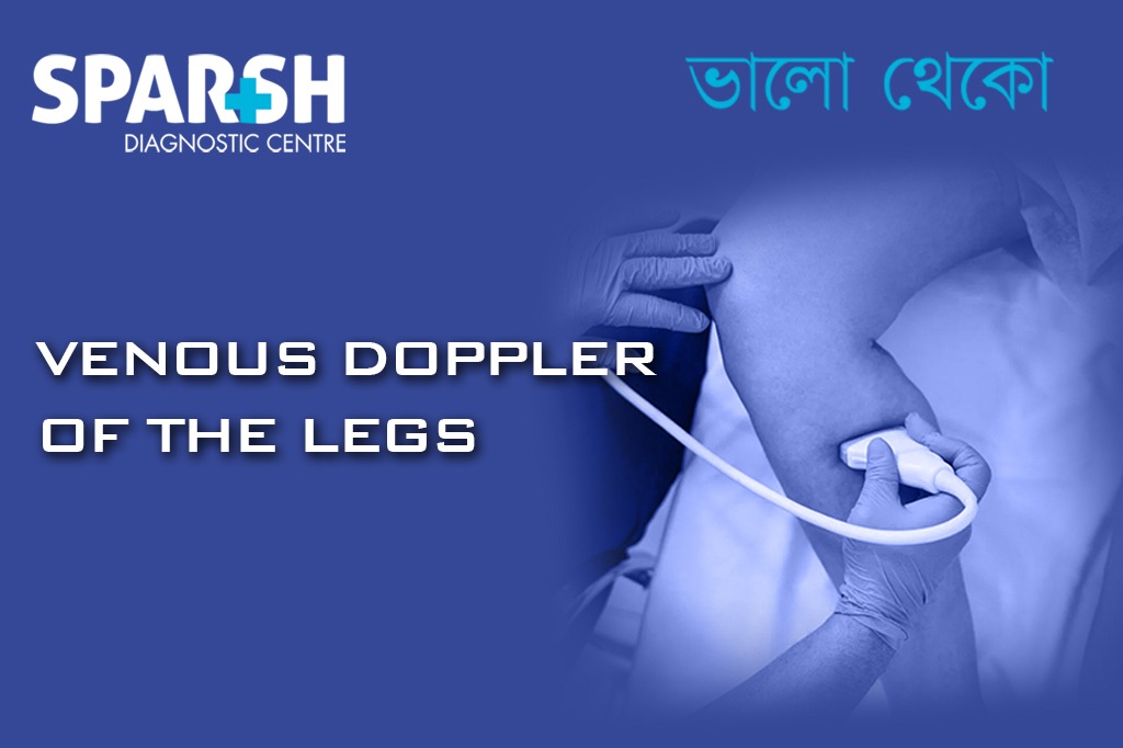
Deep Vein Thrombosis (DVT) is a serious condition that occurs when a blood clot forms in the deep veins of the legs. If left untreated, it can lead to life-threatening complications like pulmonary embolism. Among the diagnostic tools available, Venous Doppler Ultrasound of the legs stands out as the gold standard. This non-invasive test offers accurate, real-time imaging to detect clots and assess venous flow.
In this blog, we explore everything you need to know about Venous Doppler of the legs, its role in diagnosing DVT, the procedure, preparation, interpretation of results, and more.
What is Deep Vein Thrombosis (DVT)?
Deep Vein Thrombosis (DVT) is the formation of a thrombus (blood clot) in the deep veins, typically in the legs. The condition can affect one or both legs and may not always show symptoms. However, when symptoms do appear, they often include:
Swelling in one leg (rarely both)
Warmth in the affected leg
Red or discolored skin
A feeling of heaviness in the leg
The greatest danger of DVT is that the clot may dislodge and travel to the lungs, causing a pulmonary embolism (PE)—a medical emergency that can be fatal.
What is Venous Doppler of the Legs?
Venous Doppler ultrasound, also known as venous duplex ultrasonography, is a non-invasive imaging test used to evaluate blood flow in the veins of the legs. It combines traditional ultrasound with Doppler technology, which measures the direction and speed of blood flow.
The test is especially useful for detecting:
Blood clots (DVT)
Venous insufficiency
Venous reflux disorders
Obstructions or narrowed veins
Why is Venous Doppler Performed?
The primary reason for performing a Venous Doppler ultrasound of the legs is to detect or rule out DVT. It is often ordered when patients present with symptoms like:
Unexplained leg swelling
Leg pain not caused by injury
Visible varicose veins
Recent surgery, immobility, or hospitalization
A known history of clotting disorders
Family history of thrombosis
Additionally, it may be used to:
Monitor known DVT
Assess vein function before surgery
Evaluate the success of previous DVT treatment
Investigate causes of leg ulcers or chronic swelling
How Does the Doppler Ultrasound Work?
The test uses high-frequency sound waves to create images of the leg veins. A Doppler probe measures the movement of blood within the veins, helping to detect abnormalities like clots or sluggish blood flow.
Here’s what it captures:
Flow direction – whether blood is moving toward or away from the heart
Flow velocity – how fast the blood is moving
Compression response – normal veins collapse when pressure is applied; clotted veins do not
Who Needs a Venous Doppler of the Legs?
Venous Doppler is recommended for individuals who are:
Post-surgical or recently hospitalized
Pregnant, especially in the third trimester
Using hormone therapy (e.g., oral contraceptives)
Overweight or obese
Smokers
Diagnosed with cancer
Long-distance travelers (immobile for extended periods)
Experiencing unexplained leg symptoms
The test is also commonly used in emergency rooms to rule out DVT quickly.
How to Prepare for a Venous Doppler Test?
The Venous Doppler test typically does not require any special preparation. However, here are a few things to keep in mind:
Wear loose-fitting clothes for easy access to the legs.
No fasting is required.
Inform your doctor if you’re taking blood thinners or have any implants in your legs.
If you’re hospitalized, the test may be done at the bedside.
Procedure: What to Expect During the Test
The test is painless and usually takes 30 to 45 minutes.
Step-by-step overview:
Positioning: You’ll lie on your back or stomach on an examination table.
Application of gel: A water-based gel is applied to the legs to improve sound wave transmission.
Ultrasound probe: The technician moves a handheld transducer over your legs to capture images.
Compression test: Gentle pressure is applied with the probe to check vein compressibility.
Flow assessment: Doppler is used to listen to the blood flow within the veins.
Completion: Gel is wiped off and you may resume normal activities.
There is no radiation exposure, and the test is safe for pregnant women.
Venous Doppler vs. Other Tests for DVT
| Test | Invasiveness | Usefulness for DVT | Comments |
|---|---|---|---|
| Venous Doppler | Non-invasive | Gold standard | Widely used for first-line diagnosis |
| D-dimer test | Blood test | Useful for ruling out DVT | Not diagnostic alone |
| CT Venography | Invasive, with contrast | High sensitivity | Used if Doppler is inconclusive |
| MRI Venography | Non-invasive | Detailed imaging | Costly, not first-line |
How Accurate is the Venous Doppler Test?
The sensitivity and specificity of Venous Doppler for detecting proximal DVT (thigh veins) is approximately 95-98%. For distal DVT (below the knee), the accuracy is slightly lower, around 60-70%.
Factors that influence test accuracy include:
Technician experience
Patient body type
Quality of equipment
Presence of chronic venous changes
Despite its limitations, Doppler ultrasound remains the first-line test due to its reliability, cost-effectiveness, and non-invasive nature.
Interpreting the Results
Normal Result:
Veins compress easily
Blood flows smoothly without obstruction
No visible clot or venous abnormality
Abnormal Result:
Vein does not compress, suggesting a clot
Altered blood flow pattern
Venous dilation or reflux observed
A positive result confirms DVT and usually leads to immediate treatment. A negative result, especially in high-risk cases, may be followed by further testing like repeat Doppler in 5–7 days or additional imaging.
Treatment Implications After a Positive Test
Once DVT is confirmed, prompt treatment is essential to prevent complications like pulmonary embolism or post-thrombotic syndrome. Treatment options include:
Anticoagulants (blood thinners): Heparin, warfarin, or newer oral agents like rivaroxaban
Compression stockings: Improve blood flow and reduce swelling
Thrombolytic therapy: Used in severe or extensive DVT
IVC filters: Placed in patients who cannot take blood thinners
The choice of treatment depends on the location, size of clot, risk factors, and overall health of the patient.
Risks and Limitations of the Test
Venous Doppler is extremely safe, but some limitations include:
Reduced accuracy in obese patients
Difficulty in detecting small or distal clots
Variability depending on operator skill
False negatives in early-stage clots
Still, the benefits far outweigh the risks, especially given the test’s non-invasive nature.
Follow-Up and Monitoring
For patients diagnosed with DVT:
Repeat Doppler may be done to assess clot resolution or progression.
Patients on anticoagulants undergo regular monitoring of clotting status (especially with warfarin).
Long-term management may involve lifestyle changes, physical activity, and monitoring for recurrence.
Preventing DVT: When to Consider Doppler Screening
If you’re at high risk for DVT but asymptomatic, your doctor may still recommend a Doppler ultrasound, especially:
Before major orthopedic or cancer surgeries
During high-risk pregnancies
If you have a known genetic clotting disorder
Early detection can prevent severe complications and improve outcomes significantly.
Venous Doppler ultrasound of the legs is an indispensable tool in diagnosing Deep Vein Thrombosis. Quick, painless, and highly accurate, it plays a critical role in identifying clots and preventing life-threatening complications like pulmonary embolism.
If you or someone you know is experiencing symptoms of leg swelling, pain, or tenderness—especially after surgery, travel, or immobilization—don’t delay. Early testing with Doppler ultrasound can save lives.
Frequently Asked Questions (FAQs)
1. Is a Venous Doppler painful?
No, the procedure is completely painless and non-invasive.
2. How long does the test take?
Usually between 30 to 45 minutes.
3. Can I walk after a Doppler test?
Yes, you can resume all normal activities immediately after the test.
4. What if my Doppler test is negative, but I still have symptoms?
Your doctor may repeat the test after a few days or order additional tests like a D-dimer or CT scan.
Book Your Doppler Ultrasound Today at Sparsh Diagnostic Centre
At Sparsh Diagnostic Centre, we use state-of-the-art Doppler technology to deliver fast, accurate, and reliable diagnostics. Our expert radiologists ensure you receive the best possible care for your health.
📞 Call: 9830117733 / 8335049501
🌐 Visit: www.sparshdiagnostica.com
#BhaloTheko
Disclaimer:
No content on this site, regardless of date, should ever be used as a substitute for direct medical advice from your doctor or other qualified clinician.
![]()


