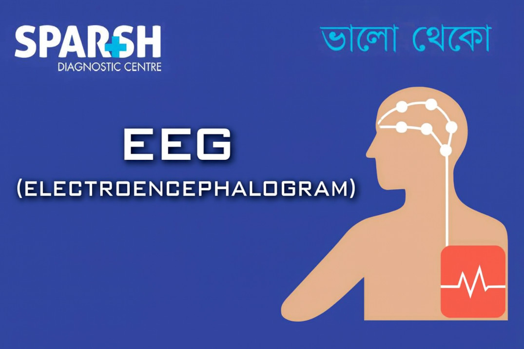
An Electroencephalogram (EEG) is one of the most widely used and reliable diagnostic tests in neurology. It measures electrical activity in the brain through small sensors placed on the scalp. These tiny electrical signals reveal crucial information about how the brain is functioning, helping doctors diagnose conditions like epilepsy, seizures, sleep disorders, encephalopathies, and more.
In today’s healthcare environment—where early diagnosis and neurological evaluation are critical—EEG plays a vital role. Whether you’re a patient preparing for an EEG, a caregiver researching treatment options, or a healthcare provider looking for patient-friendly content, this comprehensive guide will help you understand everything about the Electroencephalogram test.
What Is an EEG?
An Electroencephalogram (EEG) is a non-invasive test that records the brain’s electrical activity. The brain constantly produces tiny electrical impulses as nerve cells communicate. These signals form distinct brain wave patterns, which can be easily captured using electrodes placed on the scalp.
The EEG machine records these impulses and converts them into waveforms displayed on a screen. Neurologists study these patterns to identify abnormalities or disruptions in brain function.
Key Features of an EEG
Completely safe and painless
No radiation exposure
Can be done on patients of all ages
Widely used for diagnosing neurological disorders
Helps assess brain activity during wakefulness and sleep
Why Is an EEG Done?
Doctors recommend an EEG when they suspect a neurological issue affecting the brain’s electrical activity. Some common reasons include:
1. Epilepsy and Seizure Disorders
EEG is the gold-standard diagnostic test for epilepsy. It detects:
Abnormal electrical discharges
Risk of future seizures
Type and origin of epileptic activity
2. Sleep Disorders
Sleep-related problems often originate in the brain. EEG helps diagnose:
REM disturbances
Parasomnias
During a polysomnography (sleep study), EEG is a major component.
3. Brain Infections
EEG assists in evaluating brain activity in:
Cerebral abscess
It helps understand the severity and progression of infection.
4. Brain Tumours
Tumours—benign or malignant—can change electrical patterns. EEG helps identify:
Abnormal waveforms
Focal disturbances
Effects on surrounding brain tissue
5. Stroke and Brain Injury
After trauma or stroke, EEG can detect:
Brain swelling
Reduced electrical activity
Areas of damaged brain tissue
6. Unexplained Fainting or Confusion
Episodes of:
Loss of consciousness
Memory gaps
Disorientation
may require an EEG to determine if they are neurological in origin.
7. Assessing Brain Activity in Coma
EEG is often used in ICUs to:
Monitor brain function
Detect silent seizures
Predict recovery outcomes
How Does an EEG Work?
EEG uses electrodes (small metal discs) that are attached to the scalp using a special gel. These electrodes:
Detect electrical signals produced by brain cells
Transmit them to a computer
Display waveforms (alpha, beta, theta, delta waves)
Allow neurologists to analyse patterns
Each type of brain wave corresponds to specific mental states such as sleep, relaxation, alertness, or deep concentration.
Types of EEG Tests
There are several types of EEGs, each useful for different conditions:
1. Routine EEG
Duration: 20–40 minutes
Commonly used for diagnosing seizures, headaches, and fainting spells
2. Sleep-Deprived EEG
The patient is kept awake the night before
Helps reveal hidden seizure activity
3. Ambulatory EEG
Portable device worn for 24–72 hours
Records brain activity during daily activities
Useful for long-term monitoring
4. Video EEG Monitoring
Combines EEG with continuous video recording
Helps correlate physical symptoms with brain wave changes
Widely used in epilepsy evaluation
5. ICU EEG Monitoring
Continuous monitoring in critical care settings
Detects subclinical (silent) seizures
How to Prepare for an EEG
1. Wash Hair Before the Test
Keep hair clean and free of oils, sprays, or gels
Helps electrodes stick properly
2. Avoid Caffeine 8–12 Hours Before
Coffee, tea, and energy drinks can affect brain activity.
3. Take Regular Medication Unless Told Otherwise
Certain drugs (like anti-seizure or sedatives) may be adjusted before the test, but only under medical advice.
4. For Sleep-Deprived EEG
Stay awake the night before or sleep fewer hours as instructed.
What Happens During an EEG Test?
The EEG procedure is simple and comfortable. Here’s what to expect:
Step-by-Step Process
Electrode Placement
The technician measures your head and places electrodes using a gel.Relaxing or Lying Still
You may be asked to:Close your eyes
Breathe deeply
Look at flashing lights (photostimulation)
Recording Brain Waves
The machine records electrical activity for 20–60 minutes.Post-Test Cleaning
The gel is wiped off; you can return to normal activities immediately.
Is an EEG Painful?
No. EEG is non-invasive and painless. You won’t feel any electrical activity.
Understanding EEG Results
A neurologist interprets EEG results by analysing brain wave patterns.
Normal EEG Findings
Regular, symmetrical waveforms
Clear transitions between wakefulness and sleep
No spikes or sharp waves
Abnormal EEG Findings
EEG may show:
Spikes
Slow waves
Sharp waves
Seizure discharges
Focal abnormalities
These results help diagnose:
Epilepsy
Brain infections
Tumours
Memory disorders
Sleep disturbances
Brain injury
How Long Do EEG Results Take?
Typically 24–48 hours, depending on analysis complexity.
Risks and Safety of EEG
EEG is one of the safest diagnostic tests.
Common Concerns
No electricity enters the body
No radiation
Safe for children and pregnant women
No side effects
Rarely, flashing lights may trigger seizures in patients with photosensitive epilepsy—but medical staff are trained for immediate management.
Benefits of an EEG
Helps diagnose neurological conditions early
Guides treatment plans for seizures
Monitors brain activity in ICU
Identifies causes of confusion and fainting
Supports long-term epilepsy management
Essential in pre-surgical evaluation
EEG vs MRI: What’s the Difference?
Though both assess brain health, they serve different purposes.
| Feature | EEG | MRI |
|---|---|---|
| Function | Measures electrical activity | Shows brain structure |
| Use | Seizures, sleep disorders | Tumours, stroke, injury |
| Procedure | Electrodes on scalp | Magnetic imaging |
| Invasiveness | Non-invasive | Non-invasive |
| Duration | 20–40 mins | 15–45 mins |
Doctors often recommend both for accurate diagnosis.
After the EEG: What to Expect
You can resume daily activities immediately.
However:
Hair may feel sticky due to gel
If sedatives were used, avoid driving
Your doctor will discuss findings and recommend further tests or treatment if needed.
FAQ Section: EEG (Electroencephalogram)
1. Is EEG painful?
No, EEG is completely painless and safe.
2. How long does an EEG take?
A routine EEG usually takes 20–40 minutes. Specialized EEGs may take several hours.
3. Can I eat before an EEG?
Yes, but avoid caffeine for 8–12 hours before the test.
4. Is EEG safe for children?
Absolutely. EEG is commonly done on infants and children.
5. Can EEG detect anxiety or depression?
EEG can show brain wave patterns, but it is not used to diagnose psychiatric disorders.
6. Can I wash my hair after the test?
Yes. You may want to wash off the gel used during the procedure.
7. Do I need someone to accompany me?
Only if you’re undergoing a sleep-deprived EEG or taking sedatives.
8. Can EEG detect brain death?
Yes. EEG is often used in ICUs to assess brain function in coma patients.
An Electroencephalogram (EEG) is an essential tool in modern neurology. It helps diagnose a wide range of neurological disorders quickly, safely, and accurately. Understanding what happens during the test, why it’s needed, and how to prepare can reduce anxiety and ensure smooth testing.
#BhaloTheko
Disclaimer:
No content on this site, regardless of date, should ever be used as a substitute for direct medical advice from your doctor or other qualified clinician.

![]()